-
 Australia
-
 Austria
-
 Belgium
-
 Brazil
-
 Canada
-
 China
China
-
 Czech Republic
-
 Denmark
-
 Finland
-
 France
-
 Germany
-
 Greece
-
 Hong Kong
-
 Hungary
-
 Iceland
-
 India
India
-
 Ireland
-
 Israel
-
 Italy
Italy
-
 Japan
Japan
-
 Korea
Korea
-
 Luxembourg
-
 Malaysia
-
 Netherlands
-
 New Zealand
-
 Norway
-
 Poland
-
 Qatar
Qatar
-
 Romania
-
 Saudi Arabia
-
 Singapore
-
 Spain
Spain
-
 Sweden
-
 Switzerland
-
 Taiwan
-
 Turkey
-
 United Kingdom
-
 United States
research use only
DNA Ligase IV/LIG4 Antibody [H22J17]
Cat.No.: F7967
Application:
Reactivity:
-
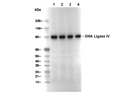 Lane 1: MOLT-4, Lane 2: MCF7, Lane 3: Hela, Lane 4: HT-1080
Usage Information
| Dilution |
|---|
|
| Application |
|---|
| WB |
| Reactivity |
|---|
| Human |
| Source |
|---|
| Rabbit Monoclonal Antibody |
| Storage Buffer |
|---|
| PBS, pH 7.2+50% Glycerol+0.05% BSA+0.01% NaN3 |
| Storage (from the date of receipt) |
|---|
| -20°C (avoid freeze-thaw cycles), 2 years |
| Predicted MW |
|---|
| 100 kDa |
| Positive Control | MOLT-4 cells; MCF7 cells; HeLa cells; HT-1080 cells |
|---|---|
| Negative Control |
Experimental Methods
| WB |
|---|
Experimental Protocol:
Sample preparation
1. Tissue: Lyse the tissue sample by adding an appropriate volume of ice-cold RIPA/Nuclear Lysis Buffer (containing Protease Inhibitor Cocktail),and homogenize the tissue at a low temperature. 2. Adherent cell: Aspirate the culture medium and wash the cells with ice-cold PBS twice. Lyse the cells by adding an appropriate volume of RIPA/Nuclear Lysis Buffer (containing Protease Inhibitor Cocktail) and put the sample on ice for 5 min. 3. Suspension cell: Transfer the culture medium to a pre-cooled centrifuge tube. Centrifuge and aspirate the supernatant. Wash the cells with ice-cold PBS twice. Lyse the cells by adding an appropriate volume of RIPA/Nuclear Lysis Buffer (containing Protease Inhibitor Cocktail) and put the sample on ice for 5 min. 4. Place the lysate into a pre-cooled microcentrifuge tube. Centrifuge at 4°C for 15 min. Collect the supernatant;
5. Remove a small volume of lysate to determine the protein concentration;
6. Combine the lysate with protein loading buffer. Boil 20 µL sample under 95-100°C for 5 min. Centrifuge for 5 min after cool down on ice.
Electrophoretic separation
1. According to the concentration of extracted protein, load appropriate amount of protein sample and marker onto SDS-PAGE gels for electrophoresis. Recommended separating gel (lower gel) concentration: 10%. Reference Table for Selecting SDS-PAGE Separation Gel Concentrations 2. Power up 80V for 30 minutes. Then the power supply is adjusted (110 V~150 V), the Marker is observed, and the electrophoresis can be stopped when the indicator band of the predyed protein Marker where the protein is located is properly separated. (Note that the current should not be too large when electrophoresis, too large current (more than 150 mA) will cause the temperature to rise, affecting the result of running glue. If high currents cannot be avoided, an ice bath can be used to cool the bath.)
Transfer membrane
1. Take out the converter, soak the clip and consumables in the pre-cooled converter;
2. Activate PVDF membrane with methanol for 1 min and rinse with transfer buffer;
3. Install it in the order of "black edge of clip - sponge - filter paper - filter paper - glue -PVDF membrane - filter paper - filter paper - sponge - white edge of clip"; 4. The protein was electrotransferred to PVDF membrane. ( 0.45 µm PVDF membrane is recommended ) Reference Table for Selecting PVDF Membrane Pore Size Specifications Recommended conditions for wet transfer: 200 mA, 120 min. ( Note that the transfer conditions can be adjusted according to the protein size. For high-molecular-weight proteins, a higher current and longer transfer time are recommended. However, ensure that the transfer tank remains at a low temperature to prevent gel melting.)
Block
1. After electrotransfer, wash the film with TBST at room temperature for 5 minutes;
2. Incubate the film in the blocking solution for 1 hour at room temperature;
3. Wash the film with TBST for 3 times, 5 minutes each time.
Antibody incubation
1. Use 5% skim milk powder to prepare the primary antibody working liquid (recommended dilution ratio for primary antibody 1:1000), gently shake and incubate with the film at 4°C overnight; 2. Wash the film with TBST 3 times, 5 minutes each time;
3. Add the secondary antibody to the blocking solution and incubate with the film gently at room temperature for 1 hour;
4. After incubation, wash the film with TBST 3 times for 5 minutes each time.
Antibody staining
1. Add the prepared ECL luminescent substrate (or select other color developing substrate according to the second antibody) and mix evenly;
2. Incubate with the film for 1 minute, remove excess substrate (keep the film moist), wrap with plastic film, and expose in the imaging system. |
Biological Description
| Specificity |
|---|
| DNA Ligase IV/LIG4 Antibody [H22J17] detects endogenous levels of total DNA Ligase IV/LIG4 protein. |
| Subcellular Location |
|---|
| Nucleus |
| Uniprot ID |
|---|
| P49917 |
| Clone |
|---|
| H22J17 |
| Synonym(s) |
|---|
| DNA ligase 4; DNA ligase IV; Polydeoxyribonucleotide synthase [ATP] 4; LIG4 |
| Background |
|---|
| DNA Ligase IV, or LIG4, is an ATP-dependent DNA ligase from the DNA ligase family that acts as the catalytic core of the non-homologous end-joining pathway, carrying out the final nick-sealing step to repair DNA double-strand breaks caused by ionizing radiation or replication stress. This enzyme contains a DNA-binding domain, a nucleotidyltransferase domain, and an oligonucleotide-binding domain that together dynamically encircle duplex DNA substrates; its active site residues, lysine 305 and lysine 471, form lysyl-AMP intermediates and coordinate phosphoanhydride bonds for 5'-phosphate adenylation, followed by nucleophilic attack from the 3'-hydroxyl group to complete ligation. LIG4's main function is to be recruited in an XRCC4 and XLF-dependent manner to double-strand break ends bound by Ku70/80, where it forms short-range synaptic complexes with a single LIG4-XRCC4 dimer bridging compatible ends for accurate ligation before any end-processing, thereby ensuring high-fidelity non-homologous end joining throughout the cell cycle and preventing the use of error-prone alternative end-joining or microhomology pathways. Its ability to bind DNA ends through both the DNA-binding domain and dynamic weak interactions stabilizes synapsis, supports ligase III as a backup when LIG4 is inactive, and is essential for V(D)J recombination in immune system development. Mutations that reduce LIG4 activity cause LIG4 syndrome, characterized by immunodeficiency, microcephaly, and radiosensitivity due to defective double-strand break rejoining, while oncogenic alterations in LIG4 can drive cancer by promoting genomic instability. |
| References |
|---|
|
Tech Support
Tel: +1-832-582-8158 Ext:3
If you have any other enquiries, please leave a message.
Products are for research use only. Not for human use. We do not sell to patients.
©Copyright 2013 Selleck Chemicals. All Rights Reserved.