
-
 Australia
-
 Austria
-
 Belgium
-
 Brazil
-
 Canada
-
 China
China
-
 Czech Republic
-
 Denmark
-
 Finland
-
 France
-
 Germany
-
 Greece
-
 Hong Kong
-
 Hungary
-
 Iceland
-
 India
India
-
 Ireland
-
 Israel
-
 Italy
Italy
-
 Japan
Japan
-
 Korea
Korea
-
 Luxembourg
-
 Malaysia
-
 Netherlands
-
 New Zealand
-
 Norway
-
 Poland
-
 Qatar
Qatar
-
 Romania
-
 Saudi Arabia
-
 Singapore
-
 Spain
Spain
-
 Sweden
-
 Switzerland
-
 Taiwan
-
 Turkey
-
 United Kingdom
-
 United States
research use only
DAG Lipase α Antibody (Rabbit mAb) [D2E6]
CatNo: F8229
Application:
Reactivity:
-
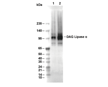 Lane 1: Mouse brain, Lane 2: Mouse cerebellum
Experiment Essentials
WB
Recommended SDS-PAGE separating gel concentration: 5%.
Recommended SDS-PAGE separating gel concentration: 5%.
Usage Information
| Dilution |
|---|
|
| Application |
|---|
| WB, IP |
| Reactivity |
|---|
| Human, Mouse, Rat |
| Source |
|---|
| Rabbit Monoclonal Antibody |
| Storage Buffer |
|---|
| PBS, pH 7.2+50% Glycerol+0.05% BSA+0.01% NaN3 |
| Storage (from the date of receipt) |
|---|
| -20°C (avoid freeze-thaw cycles), 2 years |
| Predicted MW |
|---|
| 115 kDa |
| Positive Control | Mouse brain; Mouse Cerebellum; Rat brain; Human cortex; Human cerebellum |
|---|---|
| Negative Control |
Experimental Methods
| WB |
|---|
Experimental Protocol:
Sample preparation
1. Tissue: Lyse the tissue sample by adding an appropriate volume of ice-cold RIPA/NP-40 Lysis Buffer (containing Protease Inhibitor Cocktail),and homogenize the tissue at a low temperature or lyse it by sonication on ice, then incubate on ice for 30 minutes. 2. Adherent cell: Aspirate the culture medium and wash the cells with ice-cold PBS twice. Lyse the cells by adding an appropriate volume of RIPA/NP-40 Lysis Buffer (containing Protease Inhibitor Cocktail) , sonicate to lyse the cells, and incubate on ice for 30 minutes. 3. Suspension cell: Transfer the culture medium to a pre-cooled centrifuge tube. Centrifuge and aspirate the supernatant. Wash the cells with ice-cold PBS twice. Lyse the cells by adding an appropriate volume of RIPA/NP-40 Lysis Buffer (containing Protease Inhibitor Cocktail) , sonicate to lyse the cells, and incubate on ice for 30 minutes. 4. Place the lysate into a pre-cooled microcentrifuge tube. Centrifuge at 4°C for 15 min. Collect the supernatant;
5. Remove a small volume of lysate to determine the protein concentration;
6. Combine the lysate with protein loading buffer. Boil 20 µL sample under 95-100°C for 5 min. Centrifuge for 5 min after cool down on ice.
Electrophoretic separation
1. According to the concentration of extracted protein, load appropriate amount of protein sample and marker onto SDS-PAGE gels for electrophoresis. Recommended separating gel (lower gel) concentration: 5%. Reference Table for Selecting SDS-PAGE Separation Gel Concentrations 2. Power up 80V for 30 minutes. Then the power supply is adjusted (110 V~150 V), the Marker is observed, and the electrophoresis can be stopped when the indicator band of the predyed protein Marker where the protein is located is properly separated. (Note that the current should not be too large when electrophoresis, too large current (more than 150 mA) will cause the temperature to rise, affecting the result of running glue. If high currents cannot be avoided, an ice bath can be used to cool the bath.)
Transfer membrane
1. Take out the converter, soak the clip and consumables in the pre-cooled converter;
2. Activate PVDF membrane with methanol for 1 min and rinse with transfer buffer;
3. Install it in the order of "black edge of clip - sponge - filter paper - filter paper - glue -PVDF membrane - filter paper - filter paper - sponge - white edge of clip"; 4. The protein was electrotransferred to PVDF membrane. ( 0.45 µm PVDF membrane is recommended ) Reference Table for Selecting PVDF Membrane Pore Size Specifications Recommended conditions for wet transfer: 200 mA, 120 min. ( Note that the transfer conditions can be adjusted according to the protein size. For high-molecular-weight proteins, a higher current and longer transfer time are recommended. However, ensure that the transfer tank remains at a low temperature to prevent gel melting.)
Block
1. After electrotransfer, wash the film with TBST at room temperature for 5 minutes;
2. Incubate the film in the blocking solution for 1 hour at room temperature;
3. Wash the film with TBST for 3 times, 5 minutes each time.
Antibody incubation
1. Use 5% skim milk powder to prepare the primary antibody working liquid (recommended dilution ratio for primary antibody 1:1000), gently shake and incubate with the film at 4°C overnight; 2. Wash the film with TBST 3 times, 5 minutes each time;
3. Add the secondary antibody to the blocking solution and incubate with the film gently at room temperature for 1 hour;
4. After incubation, wash the film with TBST 3 times for 5 minutes each time.
Antibody staining
1. Add the prepared ECL luminescent substrate (or select other color developing substrate according to the second antibody) and mix evenly;
2. Incubate with the film for 1 minute, remove excess substrate (keep the film moist), wrap with plastic film, and expose in the imaging system. |
Biological Description
| Specificity |
|---|
| DAG Lipase α Antibody (Rabbit mAb) [D2E6] detects endogenous levels of total DAG Lipase α protein. |
| Subcellular Location |
|---|
| Cell membrane, Cell projection, Endosome, Membrane, Postsynaptic cell membrane, Synapse |
| Uniprot ID |
|---|
| Q9Y4D2 |
| Clone |
|---|
| D2E6 |
| Synonym(s) |
|---|
| C11orf11; DAGL-alpha; DAGL(ALPHA); DAGLA; DAGLALPHA; DGL-alpha; DGLA; KIAA0659; Neural stem cell-derived dendrite regulator; NSDDR; Sn1-specific diacylglycerol lipase alpha |
| Background |
|---|
| DAG lipase α is a transmembrane serine hydrolase of the diacylglycerol lipase family that serves as a principal biosynthetic enzyme for the endocannabinoid 2‑arachidonoylglycerol (2‑AG) in the central nervous system, converting sn‑1‑acyl diacylglycerol to 2‑monoacylglycerol and free fatty acid in a calcium‑dependent manner and thereby linking membrane phospholipid turnover to cannabinoid receptor signaling. The protein has a short N‑terminal cytoplasmic segment followed by four closely spaced transmembrane helices and a large C‑terminal cytosolic catalytic domain organized as an α/β hydrolase fold, with a hydrophobic active site that contains a highly conserved Ser–Asp–His catalytic triad and a regulatory loop between β‑strands 7 and 8 that functions as a lid controlling access to the active site and carries multiple phosphorylation sites implicated in activity modulation. DAG lipase α selectively hydrolyzes the ester bond at the sn‑1 position of diacylglycerol, generating 2‑monoacylglycerol species such as 2‑AG; mechanistic studies describe a two‑step acyl‑enzyme process in which histidine‑activated serine attacks the acyl carbonyl to form a tetrahedral intermediate and monoacylglycerol product, followed by water‑mediated hydrolysis of the acyl‑enzyme to release the free fatty acid and regenerate the catalytic serine. In neuronal circuits, DAG lipase α is enriched postsynaptically on somatodendritic membranes and in perisynaptic spine regions, where it couples Gq‑linked receptor activation and depolarization to on‑demand 2‑AG synthesis for retrograde signaling; precise subcellular mapping in striatal medium spiny neurons shows co‑distribution of DAG lipase α with mGluR5 in perisynaptic zones, and DAGLα‑dependent 2‑AG mediates depolarization‑induced suppression of inhibition and excitation by activating presynaptic CB1 receptors on GABAergic and glutamatergic terminals. Genetic manipulation of DAG lipase α confirms its central role in CNS endocannabinoid signaling: DAGLα knockout mice lack trans‑synaptic 2‑AG signaling, show impaired retrograde suppression of synaptic transmission, and exhibit altered axonal growth, synaptic plasticity and adult neurogenesis, supporting a requirement for DAGLα‑generated 2‑AG in developmental wiring and ongoing circuit modulation. Beyond synaptic modulation, DAG lipase α‑derived 2‑AG and its downstream metabolites (including arachidonic acid and prostaglandins) participate in neuroinflammation and neuroprotection; inhibition or deletion of DAGLα reduces neuroinflammatory responses and has been associated with neuroprotective effects in models of neurodegenerative disease, but extensive blockade of DAGLα also disrupts neuroplasticity and alters brain lipid composition, leading to increased anxiety, depression, seizures and other neuropsychiatric manifestations, indicating that DAGLα is a tunable but sensitive node in CNS lipid signaling. Systemic studies of DAGLα knockout mice reveal broader metabolic roles: these animals are lean, show reduced food intake and weight, and have lower fasting insulin, triglycerides and total cholesterol, suggesting that DAGLα‑dependent 2‑AG contributes to appetite regulation and metabolic homeostasis and that DAGLα inhibition may offer a route to treating obesity and metabolic syndrome while also influencing gastrointestinal motility via CB1‑linked pathways. |
| References |
|---|
|
Tech Support
Tel: +1-832-582-8158 Ext:3
If you have any other enquiries, please leave a message.

Products are for research use only. Not for human use. We do not sell to patients.
©Copyright 2013 Selleck Chemicals. All Rights Reserved.