
-
 Australia
-
 Austria
-
 Belgium
-
 Brazil
-
 Canada
-
 China
China
-
 Czech Republic
-
 Denmark
-
 Finland
-
 France
-
 Germany
-
 Greece
-
 Hong Kong
-
 Hungary
-
 Iceland
-
 India
India
-
 Ireland
-
 Israel
-
 Italy
Italy
-
 Japan
Japan
-
 Korea
Korea
-
 Luxembourg
-
 Malaysia
-
 Netherlands
-
 New Zealand
-
 Norway
-
 Poland
-
 Qatar
Qatar
-
 Romania
-
 Saudi Arabia
-
 Singapore
-
 Spain
Spain
-
 Sweden
-
 Switzerland
-
 Taiwan
-
 Turkey
-
 United Kingdom
-
 United States
research use only
Cytokeratin 3 Antibody (Mouse mAb) [N24P21]
CatNo: F2198
Application:
Reactivity:
-
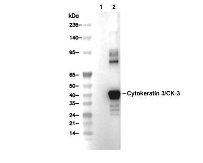 Lane 1: 293T (empty vector transfected), Lane 2: 293T (Cytokeratin full length expression vector transfected)
Usage Information
| Dilution |
|---|
|
| Application |
|---|
| IHC |
| Reactivity |
|---|
| Human |
| Source |
|---|
| Mouse Monoclonal Antibody |
| Storage Buffer |
|---|
| PBS, pH 7.2+50% Glycerol+0.05% BSA+0.01% NaN3 |
| Storage (from the date of receipt) |
|---|
| -20°C (avoid freeze-thaw cycles), 2 years |
| Predicted MW Observed MW |
|---|
| 64 kDa 65 kDa |
| *Why do the predicted and actual molecular weights differ? The following reasons may explain differences between the predicted and actual protein molecular weight. Post-translational modifications(e.g., phosphorylation, glycosylation); Splice variants and isoforms; Relative charge; Multimerization. |
| Positive Control | Human squamous papilloma of cornea tissue |
|---|---|
| Negative Control |
Experimental Methods
| IHC |
|---|
Experimental Protocol:
Deparaffinization/Rehydration
1. Deparaffinize/hydrate sections:
2. Incubate sections in three washes of xylene for 5 min each.
3. Incubate sections in two washes of 100% ethanol for 10 min each.
4. Incubate sections in two washes of 95% ethanol for 10 min each.
5. Wash sections two times in dH2O for 5 min each.
6.Antigen retrieval: For Citrate: Heat slides in a microwave submersed in 1X citrate unmasking solution until boiling is initiated; continue with 10 min at a sub-boiling temperature (95°-98°C). Cool slides on bench top for 30 min.
Staining
1. Wash sections in dH2O three times for 5 min each.
2. Incubate sections in 3% hydrogen peroxide for 10 min.
3. Wash sections in dH2O two times for 5 min each.
4. Wash sections in wash buffer for 5 min.
5. Block each section with 100–400 µl of blocking solution for 1 hr at room temperature.
6. Remove blocking solution and add 100–400 µl primary antibody diluent in to each section. Incubate overnight at 4°C.
7. Remove antibody solution and wash sections with wash buffer three times for 5 min each.
8. Cover section with 1–3 drops HRPas needed. Incubate in a humidified chamber for 30 min at room temperature.
9. Wash sections three times with wash buffer for 5 min each.
10. Add DAB Chromogen Concentrate to DAB Diluent and mix well before use.
11. Apply 100–400 µl DAB to each section and monitor closely. 1–10 min generally provides an acceptable staining intensity.
12. Immerse slides in dH2O.
13. If desired, counterstain sections with hematoxylin.
14. Wash sections in dH2O two times for 5 min each.
15. Dehydrate sections: Incubate sections in 95% ethanol two times for 10 sec each; Repeat in 100% ethanol, incubating sections two times for 10 sec each; Repeat in xylene, incubating sections two times for 10 sec each.
16. Mount sections with coverslips and mounting medium.
|
Biological Description
| Specificity |
|---|
| Cytokeratin 3 Antibody (Mouse mAb) [N24P21] detects endogenous levels of total Cytokeratin 3/CK-3 protein. |
| Subcellular Location |
|---|
| Intermediate filament, Keratin |
| Uniprot ID |
|---|
| P12035 |
| Clone |
|---|
| N24P21 |
| Synonym(s) |
|---|
| 65 kDa cytokeratin | Cytokeratin-3 | Keratin-3 | Type-II keratin Kb3 | CK-3 | K3 | KRT3 |
| Background |
|---|
| Cytokeratin 3 (CK3, gene KRT3) is a type II intermediate filament keratin that forms an obligate heteropolymeric pair with the type I keratin K12 to build the cornea-specific cytoskeletal scaffold in differentiated corneal epithelium. This network supports mechanical resilience, barrier function, and the maintenance of corneal transparency by anchoring to desmosomal and hemidesmosomal complexes and integrating with the epithelial basement membrane. CK3/K12 filaments assemble into a dense cytoplasmic lattice whose organization depends on highly conserved helix boundary motifs and the glycine-rich tail domain—regions that mediate longitudinal filament assembly and lateral filament–filament packing. Even single amino-acid substitutions in these motifs can alter filament stability and increase susceptibility to mechanical shear. Within the corneal epithelium, this keratin pair defines the differentiated suprabasal compartment, marking the transition from limbal progenitors to mature central epithelial cells and coupling cytoskeletal status to junctional complexes that stabilize epithelial sheets and preserve an optically smooth surface. Pathogenic missense mutations in KRT3, particularly those affecting the helix boundary and glycine-loop regions, disrupt heteropolymer assembly with K12, generate keratin aggregates, and weaken the intermediate filament network. This results in epithelial cell fragility, microcyst formation, and recurrent erosions characteristic of Meesmann corneal dystrophy, demonstrating how precise CK3 structure–function relationships can translate into tissue-level mechanical failure. Genetic linkage and sequencing studies show that disease-associated KRT3 variants segregate with Meesmann corneal dystrophy in affected families and are absent from unaffected controls. Different mutations in either KRT3 or KRT12 can produce a similar phenotype by compromising the same filament-stabilizing motifs, reinforcing the concept that corneal epithelial integrity depends on balanced expression and correct pairing of this keratin duo. CK3 also functions as a corneal epithelial lineage marker. Immunohistochemical detection of CK3/CK12 defines cornea-type differentiation and reveals altered expression or mislocalization of these keratins in certain corneal dystrophies. Changes in CK3 levels after chemical injury or epithelial loss correlate with shedding and re-epithelialization dynamics, linking its expression pattern to injury responses and epithelial renewal. |
| References |
|---|
|
Tech Support
Tel: +1-832-582-8158 Ext:3
If you have any other enquiries, please leave a message.

Products are for research use only. Not for human use. We do not sell to patients.
©Copyright 2013 Selleck Chemicals. All Rights Reserved.