
-
 Australia
-
 Austria
-
 Belgium
-
 Brazil
-
 Canada
-
 China
China
-
 Czech Republic
-
 Denmark
-
 Finland
-
 France
-
 Germany
-
 Greece
-
 Hong Kong
-
 Hungary
-
 Iceland
-
 India
India
-
 Ireland
-
 Israel
-
 Italy
Italy
-
 Japan
Japan
-
 Korea
Korea
-
 Luxembourg
-
 Malaysia
-
 Netherlands
-
 New Zealand
-
 Norway
-
 Poland
-
 Qatar
Qatar
-
 Romania
-
 Saudi Arabia
-
 Singapore
-
 Spain
Spain
-
 Sweden
-
 Switzerland
-
 Taiwan
-
 Turkey
-
 United Kingdom
-
 United States
research use only
Ctip1/BCL-11A Antibody (Mouse mAb) [A3G8]
CatNo: F7747
Application:
Reactivity:
-
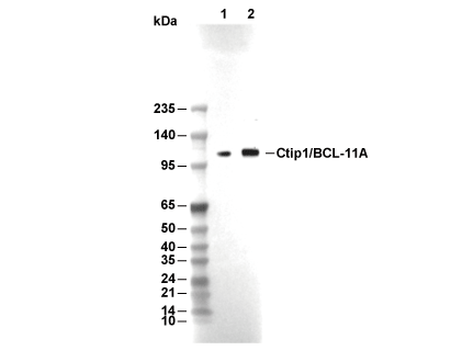 Lane 1: Jurkat, Lane 2: 293T
Experiment Essentials
WB
Exposure time of at least 60s is recommended.
Exposure time of at least 60s is recommended.
Usage Information
| Dilution |
|---|
|
| Application |
|---|
| WB, FCM |
| Reactivity |
|---|
| Mouse, Human |
| Source |
|---|
| Mouse Monoclonal Antibody |
| Storage Buffer |
|---|
| PBS, pH 7.2+50% Glycerol+0.05% BSA+0.01% NaN3 |
| Storage (from the date of receipt) |
|---|
| -20°C (avoid freeze-thaw cycles), 2 years |
| Predicted MW Observed MW |
|---|
| 91 kDa 120 kDa, 42 kDa,62 kDa |
| *Why do the predicted and actual molecular weights differ? The following reasons may explain differences between the predicted and actual protein molecular weight. Post-translational modifications(e.g., phosphorylation, glycosylation); Splice variants and isoforms; Relative charge; Multimerization. |
| Positive Control | HAP1 cells; Raji cells |
|---|---|
| Negative Control |
Experimental Methods
| WB |
|---|
Experimental Protocol:
Sample preparation
1. Tissue: Lyse the tissue sample by adding an appropriate volume of ice-cold RIPA/NP-40 Lysis Buffer (containing Protease Inhibitor Cocktail),and homogenize the tissue at a low temperature or lyse it by sonication on ice, then incubate on ice for 30 minutes. 2. Adherent cell: Aspirate the culture medium and wash the cells with ice-cold PBS twice. Lyse the cells by adding an appropriate volume of RIPA/NP-40 Lysis Buffer (containing Protease Inhibitor Cocktail) , sonicate to lyse the cells, and incubate on ice for 30 minutes. 3. Suspension cell: Transfer the culture medium to a pre-cooled centrifuge tube. Centrifuge and aspirate the supernatant. Wash the cells with ice-cold PBS twice. Lyse the cells by adding an appropriate volume of RIPA/NP-40 Lysis Buffer (containing Protease Inhibitor Cocktail) , sonicate to lyse the cells, and incubate on ice for 30 minutes. 4. Place the lysate into a pre-cooled microcentrifuge tube. Centrifuge at 4°C for 15 min. Collect the supernatant;
5. Remove a small volume of lysate to determine the protein concentration;
6. Combine the lysate with protein loading buffer. Boil 20 µL sample under 95-100°C for 5 min. Centrifuge for 5 min after cool down on ice.
Electrophoretic separation
1. According to the concentration of extracted protein, load appropriate amount of protein sample and marker onto SDS-PAGE gels for electrophoresis. Recommended separating gel (lower gel) concentration: 10%. Reference Table for Selecting SDS-PAGE Separation Gel Concentrations 2. Power up 80V for 30 minutes. Then the power supply is adjusted (110 V~150 V), the Marker is observed, and the electrophoresis can be stopped when the indicator band of the predyed protein Marker where the protein is located is properly separated. (Note that the current should not be too large when electrophoresis, too large current (more than 150 mA) will cause the temperature to rise, affecting the result of running glue. If high currents cannot be avoided, an ice bath can be used to cool the bath.)
Transfer membrane
1. Take out the converter, soak the clip and consumables in the pre-cooled converter;
2. Activate PVDF membrane with methanol for 1 min and rinse with transfer buffer;
3. Install it in the order of "black edge of clip - sponge - filter paper - filter paper - glue -PVDF membrane - filter paper - filter paper - sponge - white edge of clip"; 4. The protein was electrotransferred to PVDF membrane. ( 0.45 µm PVDF membrane is recommended ) Reference Table for Selecting PVDF Membrane Pore Size Specifications Recommended conditions for wet transfer: 200 mA, 120 min. ( Note that the transfer conditions can be adjusted according to the protein size. For high-molecular-weight proteins, a higher current and longer transfer time are recommended. However, ensure that the transfer tank remains at a low temperature to prevent gel melting.)
Block
1. After electrotransfer, wash the film with TBST at room temperature for 5 minutes;
2. Incubate the film in the blocking solution for 1 hour at room temperature;
3. Wash the film with TBST for 3 times, 5 minutes each time.
Antibody incubation
1. Use primary antibody dilution buffer to prepare the primary antibody working liquid (recommended dilution ratio for primary antibody 1:1000), gently shake and incubate with the film at 4°C overnight; 2. Wash the film with TBST 3 times, 5 minutes each time;
3. Add the secondary antibody to the blocking solution and incubate with the film gently at room temperature for 1 hour;
4. After incubation, wash the film with TBST 3 times for 5 minutes each time.
Antibody staining
1. Add the prepared ECL luminescent substrate (or select other color developing substrate according to the second antibody) and mix evenly;
2. Incubate with the film for 1 minute, remove excess substrate (keep the film moist), wrap with plastic film, and expose in the imaging system. (Exposure time of at least 60s is recommended) |
Biological Description
| Specificity |
|---|
| Ctip1/BCL-11A Antibody (Mouse mAb) [A3G8] detects endogenous levels of total Ctip1/BCL-11A protein. |
| Subcellular Location |
|---|
| Chromosome, Cytoplasm, Nucleus |
| Uniprot ID |
|---|
| Q9H165 |
| Clone |
|---|
| A3G8 |
| Synonym(s) |
|---|
| CTIP1, EVI9, KIAA1809, ZNF856, BCL11A, BCL11 transcription factor A, B-cell CLL/lymphoma 11A, Zinc finger protein 856, BCL-11A, EVI-9 |
| Background |
|---|
| CTIP1/BCL11A is a multi‑zinc‑finger transcription factor that occupies defined regulatory elements in both the β‑globin locus and the developing neocortex, where it organizes long‑range chromatin interactions and directs lineage‑specific transcriptional programs. At the human β‑globin cluster, BCL11A binds the locus control region, the ε‑globin promoter, and intergenic regions between the γ‑ and δ‑globin genes, forming a binding pattern that coincides with the fetal‑to‑adult hemoglobin switch and with a chromatin architecture in which the LCR contacts the adult β‑globin gene while interactions with γ‑globin promoters are reduced. Chromosome conformation capture and ChIP–chip data show that BCL11A participates in reconfiguring these loops and co‑occupies key sites with SOX6 and GATA1, and functional assays in adult human erythroid progenitors demonstrate that BCL11A and SOX6 cooperate to silence γ‑globin transcription, establishing BCL11A as a central component of the HbF repression complex. Genome‑wide analyses and genetic studies link erythroid BCL11A expression with HbF levels, and modulation of its dosage alters γ‑globin output in proportion to its occupancy at these regulatory elements, so BCL11A is now positioned as a primary target for HbF reactivation strategies in β‑hemoglobinopathies. In the cerebral cortex, Ctip1 is expressed by postmitotic deep‑layer projection neurons, including corticothalamic and callosal neurons, and is excluded from corticospinal motor neurons that express the related factor Ctip2, producing complementary expression domains across layer 5 and 6. Loss‑of‑function experiments show that Ctip1 is required to maintain the correct balance of deep‑layer projection neuron subtypes: deletion of Ctip1 in sensory cortex produces an excess of subcerebral projection neurons at the expense of corticothalamic and deep‑layer callosal neurons, whereas misexpression of Ctip1 represses subcerebral gene expression and their descending projections, demonstrating subtype‑specific control at the transcriptional level. Direct transcriptional targets include the cortical fate determinant Tbr1, whose repression by CTIP1 in layer 5 is necessary for acquisition of subcerebral identity, and guidance and connectivity genes such as Sema3c, which mediate neuronal polarization and radial migration of later‑born upper‑layer neurons via a defined Ctip1/Sema3c pathway. Across these systems, CTIP1/BCL11A acts through its clustered zinc fingers and corepressor‑interacting domains to bind specific promoter and enhancer sequences, assemble repressive or modulatory complexes, remodel chromatin topology, and thereby couple local transcriptional decisions to large‑scale changes in locus conformation and projection neuron wiring, linking erythroid hemoglobin switching and cortical output specification through a common transcriptional logic. |
| References |
|---|
|
Tech Support
Tel: +1-832-582-8158 Ext:3
If you have any other enquiries, please leave a message.

Products are for research use only. Not for human use. We do not sell to patients.
©Copyright 2013 Selleck Chemicals. All Rights Reserved.