
-
 Australia
-
 Austria
-
 Belgium
-
 Brazil
-
 Canada
-
 China
China
-
 Czech Republic
-
 Denmark
-
 Finland
-
 France
-
 Germany
-
 Greece
-
 Hong Kong
-
 Hungary
-
 Iceland
-
 India
India
-
 Ireland
-
 Israel
-
 Italy
Italy
-
 Japan
Japan
-
 Korea
Korea
-
 Luxembourg
-
 Malaysia
-
 Netherlands
-
 New Zealand
-
 Norway
-
 Poland
-
 Qatar
Qatar
-
 Romania
-
 Saudi Arabia
-
 Singapore
-
 Spain
Spain
-
 Sweden
-
 Switzerland
-
 Taiwan
-
 Turkey
-
 United Kingdom
-
 United States
research use only
Collagen IV Antibody (Rabbit mAb) [L21P9]
CatNo: F7452
-
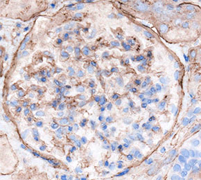 Immunohistochemical analysis of formalin fixed paraffin embedded human kidney tissue with F7452 at 1:500 dilution.
Usage Information
| Dilution |
|---|
|
| Application |
|---|
| IHC |
| Reactivity |
|---|
| Mouse, Rat, Human |
| Source |
|---|
| Rabbit Monoclonal Antibody |
| Storage Buffer |
|---|
| PBS, pH 7.2+50% Glycerol+0.05% BSA+0.01% NaN3 |
| Storage (from the date of receipt) |
|---|
| -20°C (avoid freeze-thaw cycles), 2 years |
| Predicted MW |
|---|
| 161 kDa |
| Positive Control | Human glioma tissue; Human kidney tissue; Mouse kidney tissue; Mouse cerebrum tissue; Rat kidney tissue |
|---|---|
| Negative Control |
Experimental Methods
| IHC |
|---|
Experimental Protocol:
Deparaffinization/Rehydration
1. Deparaffinize/hydrate sections:
2. Incubate sections in three washes of xylene for 5 min each.
3. Incubate sections in two washes of 100% ethanol for 10 min each.
4. Incubate sections in two washes of 95% ethanol for 10 min each.
5. Wash sections two times in dH2O for 5 min each.
6.Antigen retrieval: For Citrate: Heat slides in a microwave submersed in 1X citrate unmasking solution until boiling is initiated; continue with 10 min at a sub-boiling temperature (95°-98°C). Cool slides on bench top for 30 min.
Staining
1. Wash sections in dH2O three times for 5 min each.
2. Incubate sections in 3% hydrogen peroxide for 10 min.
3. Wash sections in dH2O two times for 5 min each.
4. Wash sections in wash buffer for 5 min.
5. Block each section with 100–400 µl of blocking solution for 1 hr at room temperature.
6. Remove blocking solution and add 100–400 µl primary antibody diluent in to each section. Incubate overnight at 4°C.
7. Remove antibody solution and wash sections with wash buffer three times for 5 min each.
8. Cover section with 1–3 drops HRPas needed. Incubate in a humidified chamber for 30 min at room temperature.
9. Wash sections three times with wash buffer for 5 min each.
10. Add DAB Chromogen Concentrate to DAB Diluent and mix well before use.
11. Apply 100–400 µl DAB to each section and monitor closely. 1–10 min generally provides an acceptable staining intensity.
12. Immerse slides in dH2O.
13. If desired, counterstain sections with hematoxylin.
14. Wash sections in dH2O two times for 5 min each.
15. Dehydrate sections: Incubate sections in 95% ethanol two times for 10 sec each; Repeat in 100% ethanol, incubating sections two times for 10 sec each; Repeat in xylene, incubating sections two times for 10 sec each.
16. Mount sections with coverslips and mounting medium.
|
Biological Description
| Specificity |
|---|
Collagen IV Antibody (Rabbit mAb) [L21P9] detects endogenous levels of total Collagen IV protein. |
| Subcellular Location |
|---|
| Basement membrane, Extracellular matrix, Secreted |
| Uniprot ID |
|---|
| P02462, P08572, P29400, P53420, Q01955 |
| Clone |
|---|
| L21P9 |
| Synonym(s) |
|---|
| Collagen alpha-1(IV) chain, COL4A1 |
| Background |
|---|
Collagen IV belongs to the non-fibrillar collagen family and is the principal structural scaffold of basement membranes, assembled not from a single alpha chain but from six distinct, genetically related alpha chains, alpha1 through alpha6, each carrying a long central collagenous domain of Gly-X-Y repeats interrupted by short noncollagenous interruptions and terminating in a noncollagenous NC1 domain. Three alpha chains associate to form a triple-helical protomer, and these protomers then oligomerize end-to-end into supramolecular networks through two defined junction points: NC1-NC1 dimerization at the carboxyl terminus of adjoining protomers, and tetramer formation through 7S domain interactions at the amino terminus, with chloride ions specifically mediating NC1 trimeric domain oligomerization to form the NC1 hexamer at the protomer-protomer interface. Chain selection into protomers and network assembly is not random but is directly encoded within the NC1 domain itself, since re-association experiments using recombinant NC1 domains show these sequences alone are sufficient to direct which alpha chains pair together, and the six chains distribute into three biologically distinct networks: an alpha1.alpha2 network that is broadly expressed across most basement membranes, an alpha3.alpha4.alpha5 network restricted to specialized tissues including the mature glomerular basement membrane, and an alpha1.alpha2.alpha5.alpha6 network found in smooth muscle basement membranes, in which an alpha5.alpha6-containing protomer links through NC1-NC1 interaction to an adjoining alpha1.alpha2 protomer. This network specificity has direct pathological consequences: the coexistence of an alpha5.alpha6 chain requirement within the same NC1-encoded assembly system explains the concomitant loss of both alpha5 and alpha6 chains from basement membranes in X-linked Alport syndrome, while in the glomerulus specifically, the normal developmental transition from a fetal alpha1.alpha2 network to the mature alpha3.alpha4.alpha5 network fails when NC1 domain mutations disrupt hexamer assembly, since formation of the alpha3.alpha4.alpha5 NC1 hexamer requires co-assembly of all three chain-specific NC1 monomers together, so a single defective chain can block network formation for the entire triplet. In Goodpasture syndrome, autoantibodies target conformational epitopes within this same alpha3.alpha4.alpha5 NC1 hexamer, directly implicating the NC1 assembly interface as both the structural determinant of network integrity and the antigenic target underlying autoimmune basement membrane disease. |
| References |
|---|
|
Tech Support
Tel: +1-832-582-8158 Ext:3
If you have any other enquiries, please leave a message.

Products are for research use only. Not for human use. We do not sell to patients.
©Copyright 2013 Selleck Chemicals. All Rights Reserved.