
-
 Argentina
-
 Australia
-
 Austria
-
 Belgium
-
 Brazil
-
 Canada
-
 China
China
-
 Czech Republic
-
 Denmark
-
 Finland
-
 France
-
 Germany
-
 Greece
-
 Hong Kong
-
 Hungary
-
 Iceland
-
 India
India
-
 Ireland
-
 Israel
-
 Italy
Italy
-
 Japan
Japan
-
 Korea
Korea
-
 Luxembourg
-
 Malaysia
-
 Netherlands
-
 New Zealand
-
 Norway
-
 Poland
-
 Portugal
-
 Qatar
Qatar
-
 Romania
-
 Saudi Arabia
-
 Singapore
-
 Spain
Spain
-
 Sweden
-
 Switzerland
-
 Taiwan
-
 Turkey
-
 United Kingdom
-
 United States
research use only
Cdc20 Antibody (Rabbit mAb) [L21M22]
CatNo: F9381
Application:
Reactivity:
-
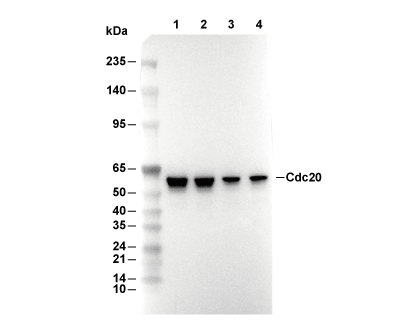 Lane 1: HeLa, Lane 2: PC3, Lane 3: DU145, Lane 4: LNCaP
Usage Information
| Dilution |
|---|
|
| Application |
|---|
| WB, IP, IHC, FCM |
| Reactivity |
|---|
| Human, Mouse, Rat |
| Source |
|---|
| Rabbit Monoclonal Antibody |
| Storage Buffer |
|---|
| PBS, pH 7.2+50% Glycerol+0.05% BSA+0.01% NaN3 |
| Storage (from the date of receipt) |
|---|
| -20°C (avoid freeze-thaw cycles), 2 years |
| Predicted MW Observed MW |
|---|
| 55 kDa 51 kDa,36 kDa |
| *Why do the predicted and actual molecular weights differ? The following reasons may explain differences between the predicted and actual protein molecular weight. Post-translational modifications(e.g., phosphorylation, glycosylation); Splice variants and isoforms; Relative charge; Multimerization. |
| Positive Control | Human ovarian carcinoma tissue; Human colon tissue; Human tonsil tissue; Rat testis tissue; Mouse testis tissue; Mouse colon tissue; Human testis tissue; Rat cerebrum tissue; Rat colon tissue; PC-3 cells; HeLa cells (Nocodazole, 100ng/ml, 16 h) |
|---|---|
| Negative Control | Human cerebrum tissue; Mouse cerebrum tissue; Rat cerebrum tissue; Human liver tissue; Mouse kidney tissue; Mouse brain tissue; Rat brain tissue; Rat liver tissue |
Experimental Methods
| WB |
|---|
Experimental Protocol:
Sample preparation
1. Tissue: Lyse the tissue sample by adding an appropriate volume of ice-cold RIPA/NP-40 Lysis Buffer (containing Protease Inhibitor Cocktail),and homogenize the tissue at a low temperature or lyse it by sonication on ice, then incubate on ice for 30 minutes. 2. Adherent cell: Aspirate the culture medium and wash the cells with ice-cold PBS twice. Lyse the cells by adding an appropriate volume of RIPA/NP-40 Lysis Buffer (containing Protease Inhibitor Cocktail) , sonicate to lyse the cells, and incubate on ice for 30 minutes. 3. Suspension cell: Transfer the culture medium to a pre-cooled centrifuge tube. Centrifuge and aspirate the supernatant. Wash the cells with ice-cold PBS twice. Lyse the cells by adding an appropriate volume of RIPA/NP-40 Lysis Buffer (containing Protease Inhibitor Cocktail) , sonicate to lyse the cells, and incubate on ice for 30 minutes. 4. Place the lysate into a pre-cooled microcentrifuge tube. Centrifuge at 4°C for 15 min. Collect the supernatant;
5. Remove a small volume of lysate to determine the protein concentration;
6. Combine the lysate with protein loading buffer. Boil 20 µL sample under 95-100°C for 5 min. Centrifuge for 5 min after cool down on ice.
Electrophoretic separation
1. According to the concentration of extracted protein, load appropriate amount of protein sample and marker onto SDS-PAGE gels for electrophoresis. Recommended separating gel (lower gel) concentration: 10%. Reference Table for Selecting SDS-PAGE Separation Gel Concentrations 2. Power up 80V for 30 minutes. Then the power supply is adjusted (110 V~150 V), the Marker is observed, and the electrophoresis can be stopped when the indicator band of the predyed protein Marker where the protein is located is properly separated. (Note that the current should not be too large when electrophoresis, too large current (more than 150 mA) will cause the temperature to rise, affecting the result of running glue. If high currents cannot be avoided, an ice bath can be used to cool the bath.)
Transfer membrane
1. Take out the converter, soak the clip and consumables in the pre-cooled converter;
2. Activate PVDF membrane with methanol for 1 min and rinse with transfer buffer;
3. Install it in the order of "black edge of clip - sponge - filter paper - filter paper - glue -PVDF membrane - filter paper - filter paper - sponge - white edge of clip"; 4. The protein was electrotransferred to PVDF membrane. ( 0.45 µm PVDF membrane is recommended ) Reference Table for Selecting PVDF Membrane Pore Size Specifications Recommended conditions for wet transfer: 200 mA, 120 min. ( Note that the transfer conditions can be adjusted according to the protein size. For high-molecular-weight proteins, a higher current and longer transfer time are recommended. However, ensure that the transfer tank remains at a low temperature to prevent gel melting.)
Block
1. After electrotransfer, wash the film with TBST at room temperature for 5 minutes;
2. Incubate the film in the blocking solution for 1 hour at room temperature;
3. Wash the film with TBST for 3 times, 5 minutes each time.
Antibody incubation
1. Use primary antibody dilution buffer to prepare the primary antibody working liquid (recommended dilution ratio for primary antibody 1:1000), gently shake and incubate with the film at 4°C overnight; 2. Wash the film with TBST 3 times, 5 minutes each time;
3. Add the secondary antibody to the blocking solution and incubate with the film gently at room temperature for 1 hour;
4. After incubation, wash the film with TBST 3 times for 5 minutes each time.
Antibody staining
1. Add the prepared ECL luminescent substrate (or select other color developing substrate according to the second antibody) and mix evenly;
2. Incubate with the film for 1 minute, remove excess substrate (keep the film moist), wrap with plastic film, and expose in the imaging system. |
| IHC |
|---|
Experimental Protocol:
Deparaffinization/Rehydration
1. Deparaffinize/hydrate sections:
2. Incubate sections in three washes of xylene for 5 min each.
3. Incubate sections in two washes of 100% ethanol for 10 min each.
4. Incubate sections in two washes of 95% ethanol for 10 min each.
5. Wash sections two times in dH2O for 5 min each.
6.Antigen retrieval: For Citrate: Heat slides in a microwave submersed in 1X citrate unmasking solution until boiling is initiated; continue with 10 min at a sub-boiling temperature (95°-98°C). Cool slides on bench top for 30 min.
Staining
1. Wash sections in dH2O three times for 5 min each.
2. Incubate sections in 3% hydrogen peroxide for 10 min.
3. Wash sections in dH2O two times for 5 min each.
4. Wash sections in wash buffer for 5 min.
5. Block each section with 100–400 µl of blocking solution for 1 hr at room temperature.
6. Remove blocking solution and add 100–400 µl primary antibody diluent in to each section. Incubate overnight at 4°C.
7. Remove antibody solution and wash sections with wash buffer three times for 5 min each.
8. Cover section with 1–3 drops HRPas needed. Incubate in a humidified chamber for 30 min at room temperature.
9. Wash sections three times with wash buffer for 5 min each.
10. Add DAB Chromogen Concentrate to DAB Diluent and mix well before use.
11. Apply 100–400 µl DAB to each section and monitor closely. 1–10 min generally provides an acceptable staining intensity.
12. Immerse slides in dH2O.
13. If desired, counterstain sections with hematoxylin.
14. Wash sections in dH2O two times for 5 min each.
15. Dehydrate sections: Incubate sections in 95% ethanol two times for 10 sec each; Repeat in 100% ethanol, incubating sections two times for 10 sec each; Repeat in xylene, incubating sections two times for 10 sec each.
16. Mount sections with coverslips and mounting medium.
|
| IP |
|---|
Experimental Procedure:
Reagents and Preparation
1. Basic buffers: 1× PBS or 1× TBS (pre-chilled on ice); 1× cell lysis buffer (add 1 mM PMSF protease inhibitor immediately before use; add phosphatase inhibitors for phosphoproteins).
2. Capture medium: Protein A/G magnetic beads or agarose beads (Sepharose beads).
3. Controls: Use an isotype control IgG matching the host species and IgG subclass of the primary antibody, at the same concentration. It is recommended to reserve a portion of the cell lysate that has not been subjected to immunoprecipitation as the Input control; if necessary, a beads-only control (beads added without antibody) may also be included.
4. Loading and elution reagents: 3× or 4× SDS sample loading buffer (containing DTT/reducing agent); if non-denaturing elution is to be performed, prepare the corresponding elution buffer and neutralization buffer separately; for denaturing elution, 1× PBS is typically used to dilute the 3× or 4× SDS sample loading buffer to 1×.
Cell Lysis and Sample Preparation (Native Protein Extraction)
1. Cell harvesting: Discard the culture medium and wash the cells once with ice-cold 1× PBS.
2. Cell lysis: Add 0.5–1 mL of ice-cold 1× cell lysis buffer to a 10-cm culture dish and incubate on ice for 5–10 minutes.
3. Lysate collection and clarification: Scrape the cells and transfer the lysate to a microcentrifuge tube. Some adherent cells may require detachment using digestive enzymes or mechanical methods. Centrifuge at 14,000 rpm at 4°C for 5–15 min. Collect the supernatant as the clarified cell lysate. It is recommended to determine the protein concentration.
Pre-clearing and Immunoprecipitation Reaction
1. Pre-clearing of the lysate (recommended): Take an appropriate amount of beads (magnetic beads or agarose beads) and pre-wash 2–3 times with cell lysis buffer or 1× TBS/PBS. After pre-washing, it is recommended to remove the wash buffer as completely as possible before adding subsequent reagents, to avoid extra dilution of the sample. For lysis systems containing detergents or specific salt concentrations, prefer pre-washing with a buffer identical or compatible with the lysis buffer to minimize buffer-system mismatches.
2. Pre-clearing treatment: Mix the cell lysate with the pre-washed beads and incubate with rotation at room temperature for 30–60 min, or at 4°C with rotation for 1–2 h.
When using agarose beads, pellet the beads by centrifugation after incubation and collect the supernatant.
When using magnetic beads, separate the beads using a magnetic stand after incubation and collect the supernatant.
This step removes proteins that bind non-specifically to the beads. For unstable proteins, phosphoproteins, or protein complexes, incubation at 4°C is preferred to minimize protein degradation, dephosphorylation, or complex dissociation. Beads used for pre-clearing should generally not be pre-coupled with a specific antibody, to avoid loss of the target antigen.
3. Formation of the “bead–antibody–antigen” immunocomplex
Either of the following two approaches may be used:
3.1 Pre-form the antibody–antigen complex, then add the beads: Add an appropriate amount of primary antibody to the pre-cleared supernatant obtained in step 2, and set up an isotype control IgG group in parallel. Incubate with rotation at 4°C overnight (recommended), or at room temperature with rotation for 2 h, to allow formation of the antibody–antigen complex.
In parallel, take an appropriate amount of beads and pre-wash them as described in step 1 of this section. Add the antibody–antigen complex to the pre-washed magnetic or agarose beads and incubate with rotation at room temperature for 30 min–1 h, or at 4°C with rotation for 1–2 h, to allow the antibody–antigen complex to bind efficiently to the beads.
3.2 Pre-form the bead–antibody complex, then add the cell lysate: Dilute the primary antibody in cell lysis buffer at the dilution ratio recommended in the antibody instruction manual to prepare the antibody working solution. Add an appropriate amount of pre-washed magnetic beads to the antibody working solution and incubate with rotation at room temperature for 15 min, or at 4°C with rotation for 1 h, to form the bead–antibody complex.
Recover the beads by magnetic separation or centrifugation, discard the supernatant, and pre-wash the beads 2–3 times with 1× TBS. Then add the cell lysate and incubate with rotation at 4°C overnight (recommended), or at room temperature with rotation for 2 h.
Washing of the Precipitate and Sample Elution
1. Once immunoprecipitation is complete, separate the beads according to their type and discard the supernatant.
Magnetic beads are separated using a magnetic stand.
For agarose beads, low-speed centrifugation (500–1000 rpm) appropriate for the bead specifications should be used, to avoid bead compaction or damage caused by high-speed centrifugation.
2. Washing the beads: Gently wash the beads 3–5 times with ice-cold 1× cell lysis buffer or 1× TBS/TBST. Keep the temperature low throughout the washes. After each addition of wash buffer, mix gently to fully resuspend the beads; then separate the beads using a magnetic stand or appropriate centrifugation, depending on the bead type, and remove the wash buffer thoroughly to minimize unbound and non-specifically bound components. During each wash, remove as much supernatant as possible while avoiding aspiration of the beads. After the final wash, remove residual wash buffer as completely as possible to prevent dilution of the eluate or interference with downstream analyses. Keep the samples on ice after each wash.
3. Sample elution (choose one of the following):
3.1 Denaturing direct elution (most commonly used): Add SDS sample loading buffer to the bead pellet to a final concentration of 1×. If 3× or 4× SDS sample loading buffer is used, it must first be diluted to 1×. Mix well and heat at 95–100°C for 5 minutes. Separate the beads and collect the supernatant for subsequent electrophoresis.
3.2 Chemical / non-denaturing elution (preserves protein activity): After the final wash, separate the beads using a magnetic stand or low-speed centrifugation, depending on the bead type, and remove residual wash buffer as completely as possible to prevent dilution of the eluate. Add an appropriate amount of acidic elution buffer / high-salt elution buffer to the beads and resuspend them thoroughly. Mix gently and incubate briefly at room temperature to allow the immunocomplex to dissociate from the beads. Separate the beads with a magnetic stand or low-speed centrifugation, and transfer the eluate to a collection tube pre-filled with an appropriate amount of neutralization buffer; adjust the pH if necessary. To improve recovery, elution may be repeated and the eluates pooled. The composition and volume of the elution buffer and neutralization buffer should be determined according to the instructions for the beads and kit used.
3.3 Post-elution sample handling
For protein activity assays, native protein complex analysis, or other non-denaturing analyses: do not add SDS sample loading buffer; keep the sample on ice and proceed to the subsequent experiment as quickly as possible.
For SDS-PAGE or Western blot: add SDS sample loading buffer to the neutralized or desalted eluate to a final concentration of 1×, and then process the sample according to the subsequent electrophoresis requirements.
Select appropriate storage conditions according to the stability of the target protein and the requirements of downstream experiments, and avoid repeated freeze–thaw cycles.
Downstream Analysis
Western blot analysis: Take the supernatant and perform SDS-PAGE electrophoresis followed by membrane transfer. It is recommended to use light- and heavy-chain-specific or conformation-specific secondary antibodies to avoid interference of the immunoglobulin heavy/light chain bands (50 kDa / 25 kDa) with detection of the target protein. It is recommended to include Input, isotype control IgG-IP, and target antibody-IP samples on the same Western blot; if necessary, a beads-only control may also be added to evaluate immunoprecipitation efficiency and non-specific binding. Kinase activity assay (if applicable): skip the denaturation step, wash the beads with kinase buffer, then add substrate and ATP directly to perform the kinase reaction.
Note: All lysis and immunoprecipitation procedures should be performed at 4°C or on ice to preserve the native conformation of proteins as much as possible and prevent their degradation. |
Biological Description
| Specificity |
|---|
| Cdc20 Antibody (Rabbit mAb) [L21M22] detects endogenous levels of total Cdc20 protein. |
| Subcellular Location |
|---|
| Centromere, Chromosome, Cytoplasm, Cytoskeleton, Kinetochore |
| Uniprot ID |
|---|
| Q12834 |
| Clone |
|---|
| L21M22 |
| Synonym(s) |
|---|
| Cell division cycle protein 20 homolog | p55CDC | CDC20 |
| Background |
|---|
| Cdc20 is an evolutionarily conserved WD40‑repeat protein that functions as the primary activator and substrate adaptor of the anaphase‑promoting complex/cyclosome (APC/C) during early mitosis, where it controls ubiquitin‑dependent degradation of key mitotic regulators and thereby coordinates metaphase–anaphase transition, chromosome segregation and mitotic exit. The protein contains an N‑terminal C box and a C‑terminal seven‑blade WD40 β‑propeller that together provide binding surfaces for APC/C, destruction motifs (D boxes and KEN boxes) in substrates such as securin and cyclin B, and regulatory factors including spindle checkpoint proteins, while additional sequence motifs such as the CRY box contribute to checkpoint signaling by modulating Cdc20’s association with inhibitory complexes. In metaphase, Cdc20 engages APC/C to form APC/C^Cdc20, an E3 ligase complex that works with E2 enzymes such as UbcH10 to polyubiquitylate securin and cyclin B, triggering separase activation and cohesin cleavage for sister chromatid separation and initiating cyclin B degradation to promote exit into anaphase and telophase; multiple mechanisms, including phosphorylation, spatial regulation and spindle assembly checkpoint (SAC) control, ensure that Cdc20‑APC/C activity is restrained until all chromosomes achieve proper bipolar attachment. The SAC converges on Cdc20 through assembly of the mitotic checkpoint complex (MCC), a heterotetramer containing MAD2, BUBR1, BUB3 and a Cdc20 molecule, which binds and inhibits APC/C^Cdc20 and prevents ubiquitination of securin and cyclin B while unattached kinetochores signal; structural and functional studies distinguish APC‑bound Cdc20 (CDC20^A) from MCC‑bound Cdc20 (CDC20^M), and clean dissection of Cdc20 motifs shows that the CRY box is critical for efficient MCC formation and checkpoint signaling, reinforcing the view that Cdc20 is the central integrator of kinetochore‑derived SAC signals into APC/C output. Beyond its canonical role at the metaphase–anaphase transition, Cdc20 has been shown to transcriptionally up‑regulate UbcH10 by physically interacting with an APC/C–CBP/p300 complex and recruiting it to the UbcH10 promoter in a cell‑cycle‑specific manner, demonstrating that Cdc20 can act as a transcriptional co‑regulator that co‑ordinates expression of its E2 partner with mitotic progression and potentially amplifies APC/C activity in proliferating cells. In cancer biology, Cdc20 is frequently overexpressed across a wide spectrum of solid tumors and hematologic malignancies, and high Cdc20 levels correlate with genomic instability and poor overall survival in many solid cancers, consistent with its role at the crossroads between chromosome segregation fidelity and cell‑cycle progression; deregulation of the Rb–E2F1 axis can up‑regulate Cdc20 and engage APC/C^Cdc20 transactivation, promoting chromosomal instability and aneuploidy. In gastric cancer and precursor lesions, immunohistochemical studies show that MAD2 and Cdc20 are most highly expressed in high‑grade dysplasia, with Cdc20 specifically staining the outer cells of cell‑in‑cell (entosis) structures and their overexpression associating with intestinal histology and favorable recurrence‑free and cancer‑specific survival, suggesting that Cdc20 levels and localization can serve both as markers of chromosomal instability–type tumors and as indicators of distinct clinicopathological behavior. Preclinical work using small‑molecule APC/C^Cdc20 inhibitors such as Apcin and proTAME demonstrates that blocking Cdc20 function induces mitotic arrest and apoptosis in lymphoma and multiple myeloma cells and synergizes with clinically relevant drugs, underscoring the therapeutic potential of targeting Cdc20–APC/C in malignancies that rely on high Cdc20 activity for unchecked cell division. |
| References |
|---|
|
Tech Support
Tel: +1-832-582-8158 Ext:3
If you have any other enquiries, please leave a message.

Products are for research use only. Not for human use. We do not sell to patients.
©Copyright 2013 Selleck Chemicals. All Rights Reserved.