-
 Australia
-
 Austria
-
 Belgium
-
 Brazil
-
 Canada
-
 China
China
-
 Czech Republic
-
 Denmark
-
 Finland
-
 France
-
 Germany
-
 Greece
-
 Hong Kong
-
 Hungary
-
 Iceland
-
 India
India
-
 Ireland
-
 Israel
-
 Italy
Italy
-
 Japan
Japan
-
 Korea
Korea
-
 Luxembourg
-
 Malaysia
-
 Netherlands
-
 New Zealand
-
 Norway
-
 Poland
-
 Qatar
Qatar
-
 Romania
-
 Saudi Arabia
-
 Singapore
-
 Spain
Spain
-
 Sweden
-
 Switzerland
-
 Taiwan
-
 Turkey
-
 United Kingdom
-
 United States
research use only
CD63 Antibody (Rabbit mAb) [P10C2]
Cat.No.: F5010
Application:
Reactivity:
-
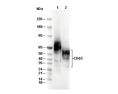 Lane 1: THP-1, Lane 2: COS-7
Experiment Essentials
WB
Recommended wet transfer conditions: 200 mA, 60 min.
Recommended wet transfer conditions: 200 mA, 60 min.
Usage Information
| Dilution |
|---|
|
| Application |
|---|
| WB |
| Reactivity |
|---|
| Human, Monkey |
| Source |
|---|
| Rabbit Monoclonal Antibody |
| Storage Buffer |
|---|
| PBS, pH 7.2+50% Glycerol+0.05% BSA+0.01% NaN3 |
| Storage (from the date of receipt) |
|---|
| -20°C (avoid freeze-thaw cycles), 2 years |
| Predicted MW |
|---|
| 25-60 kDa |
| Positive Control | THP1 cells; 293T cells; SK-MEL-28 cells; COS-7 cells |
|---|---|
| Negative Control | Jurkat cells; MOLT-4 cells |
Experimental Methods
| WB |
|---|
Experimental Protocol:
Sample preparation
1. Tissue: Lyse the tissue sample by adding an appropriate volume of ice-cold RIPA/NP-40 Lysis Buffer (containing Protease Inhibitor Cocktail),and homogenize the tissue at a low temperature or lyse it by sonication on ice, then incubate on ice for 30 minutes. 2. Adherent cell: Aspirate the culture medium and wash the cells with ice-cold PBS twice. Lyse the cells by adding an appropriate volume of RIPA/NP-40 Lysis Buffer (containing Protease Inhibitor Cocktail), sonicate to lyse the cells, and incubate on ice for 30 minutes. 3. Suspension cell: Transfer the culture medium to a pre-cooled centrifuge tube. Centrifuge and aspirate the supernatant. Wash the cells with ice-cold PBS twice. Lyse the cells by adding an appropriate volume of RIPA/NP-40 Lysis Buffer (containing Protease Inhibitor Cocktail), sonicate to lyse the cells, and incubate on ice for 30 minutes. 4. Place the lysate into a pre-cooled microcentrifuge tube. Centrifuge at 4°C for 15 min. Collect the supernatant;
5. Remove a small volume of lysate to determine the protein concentration;
6. Combine the lysate with protein loading buffer. Boil 20 µL sample under 95-100°C for 5 min. Centrifuge for 5 min after cool down on ice.
Electrophoretic separation
1. According to the concentration of extracted protein, load appropriate amount of protein sample and marker onto SDS-PAGE gels for electrophoresis. Recommended separating gel (lower gel) concentration: 10%. Reference Table for Selecting SDS-PAGE Separation Gel Concentrations 2. Power up 80V for 30 minutes. Then the power supply is adjusted (110 V~150 V), the Marker is observed, and the electrophoresis can be stopped when the indicator band of the predyed protein Marker where the protein is located is properly separated. (Note that the current should not be too large when electrophoresis, too large current (more than 150 mA) will cause the temperature to rise, affecting the result of running glue. If high currents cannot be avoided, an ice bath can be used to cool the bath.)
Transfer membrane
1. Take out the converter, soak the clip and consumables in the pre-cooled converter;
2. Activate PVDF membrane with methanol for 1 min and rinse with transfer buffer;
3. Install it in the order of "black edge of clip - sponge - filter paper - filter paper - glue -PVDF membrane - filter paper - filter paper - sponge - white edge of clip"; 4. The protein was electrotransferred to PVDF membrane. ( 0.45 µm PVDF membrane is recommended ) Reference Table for Selecting PVDF Membrane Pore Size Specifications Recommended conditions for wet transfer: 200 mA, 60 min. ( Note that the transfer conditions can be adjusted according to the protein size. For high-molecular-weight proteins, a higher current and longer transfer time are recommended. However, ensure that the transfer tank remains at a low temperature to prevent gel melting.)
Block
1. After electrotransfer, wash the film with TBST at room temperature for 5 minutes;
2. Incubate the film in the blocking solution for 1 hour at room temperature;
3. Wash the film with TBST for 3 times, 5 minutes each time.
Antibody incubation
1. Use 5% skim milk powder to prepare the primary antibody working liquid (recommended dilution ratio for primary antibody 1:1000), gently shake and incubate with the film at 4°C overnight; 2. Wash the film with TBST 3 times, 5 minutes each time;
3. Add the secondary antibody to the blocking solution and incubate with the film gently at room temperature for 1 hour;
4. After incubation, wash the film with TBST 3 times for 5 minutes each time.
Antibody staining
1. Add the prepared ECL luminescent substrate (or select other color developing substrate according to the second antibody) and mix evenly;
2. Incubate with the film for 1 minute, remove excess substrate (keep the film moist), wrap with plastic film, and expose in the imaging system. |
Biological Description
| Specificity |
|---|
| CD63 Antibody (Rabbit mAb) [P10C2] detects endogenous levels of total CD63 protein. |
| Subcellular Location |
|---|
| Cell membrane, Endosome, Lysosome, Membrane, Secreted |
| Uniprot ID |
|---|
| P08962 |
| Clone |
|---|
| P10C2 |
| Synonym(s) |
|---|
| CD63 antigen; Granulophysin; Lysosomal-associated membrane protein 3 (LAMP-3); Limp1; Melanoma-associated antigen ME491; OMA81H; Ocular melanoma-associated antigen; Tetraspanin-30 (Tspan-30); CD63; MLA1; TSPAN30 |
| Background |
|---|
| CD63, a (glycosylated) lysosome-associated membrane protein 3 (LAMP3) of the tetraspanin superfamily, is ubiquitously expressed across hematopoietic, endothelial, and melanocytic lineages, where it organizes tetraspanin-enriched microdomains (TEMs) to coordinate integrin signaling, endosomal sorting, and extracellular vesicle (EV) biogenesis. CD63 has four transmembrane domains (TM1–4) forming two tight hairpins, with a small extracellular loop 1 (ECL1, aa 40–70; conserved CCG motif disulfide) and a large ECL2 (aa 113–175; cholesterol-binding palm domain for partner lateral diffusion); cytoplasmic tails (N-term 1–22/C-term 201–237) recruit syntenin-1 and CD47 via dileucine motifs, enabling clathrin/caveolar endocytosis. ESCRT-independent multivesicular endosome (MVE) formation, CD63 scaffolds PMEL luminal domain sorting into intraluminal vesicles (ILVs) of melanosomes to initiate amyloid fibril polymerization (~4-fold fibril increase; CD63 knockdown leads to lysosomal degradation). CD63 also bridges endosomal-autophagy flux, inhibiting mTORC1 (decreasing p-mTOR and retaining LAMTOR1) and regulates EBV LMP1 secretion, balancing signal transduction (CD63 knockout increases LMP1 and mTOR hyperactivation, resulting in large autophagic vacuoles). CD63 is essential for melanosome maturation (PMEL fibrillogenesis), platelet activation (Weibel-Palade body fusion), mast cell degranulation (syntaxin-4 docking), and endothelial permeability (VEGF–integrin-αvβ3 signaling), and is an exosomal marker (~50% EV enrichment). CD63 is upregulated in melanoma, promoting metastasis via TEM–integrin αvβ3/ITGB1 trafficking; CD63 knockout disrupts lysosomal homeostasis, sensitizing cells to lysosomal inhibitors, while paradoxically acting as a tumor suppressor in colon cancer via apoptosis induction. |
| References |
|---|
|
Tech Support
Tel: +1-832-582-8158 Ext:3
If you have any other enquiries, please leave a message.
Products are for research use only. Not for human use. We do not sell to patients.
©Copyright 2013 Selleck Chemicals. All Rights Reserved.