
-
 Australia
-
 Austria
-
 Belgium
-
 Brazil
-
 Canada
-
 China
China
-
 Czech Republic
-
 Denmark
-
 Finland
-
 France
-
 Germany
-
 Greece
-
 Hong Kong
-
 Hungary
-
 Iceland
-
 India
India
-
 Ireland
-
 Israel
-
 Italy
Italy
-
 Japan
Japan
-
 Korea
Korea
-
 Luxembourg
-
 Malaysia
-
 Netherlands
-
 New Zealand
-
 Norway
-
 Poland
-
 Qatar
Qatar
-
 Romania
-
 Saudi Arabia
-
 Singapore
-
 Spain
Spain
-
 Sweden
-
 Switzerland
-
 Taiwan
-
 Turkey
-
 United Kingdom
-
 United States
research use only
CCL20/MIP-3α Antibody (Rabbit mAb) [G18F18]
CatNo: F8584
Application:
Reactivity:
-
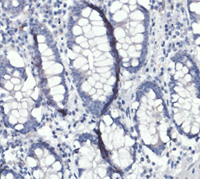 Immunohistochemical analysis of formalin fixed paraffin embedded human colon tissue with F8584 at 1:2000 dilution.
Usage Information
| Dilution |
|---|
|
| Application |
|---|
| IHC, IF, FCM |
| Reactivity |
|---|
| Human |
| Source |
|---|
| Rabbit Monoclonal Antibody |
| Storage Buffer |
|---|
| PBS, pH 7.2+50% Glycerol+0.05% BSA+0.01% NaN3 |
| Storage (from the date of receipt) |
|---|
| -20°C (avoid freeze-thaw cycles), 2 years |
| Predicted MW |
|---|
| 11 kDa |
| Positive Control | Human tonsil tissue; Human colon tissue; THP-1 cells (PMA, 100ng/ml, 56 h)(lipopolysaccharide, 1ug/ml and Brefeldin A, 300ng/ml, 16 h) |
|---|---|
| Negative Control | THP-1 cells |
Experimental Methods
| IF |
|---|
Experimental Protocol:
Sample Preparation
1. Adherent Cells: Place a clean, sterile coverslip in a culture dish. Once the cells grow to near confluence as a monolayer, remove the coverslip for further use.
2. Suspension Cells: Seed the cells onto a clean, sterile slide coated with poly-L-lysine.
3. Frozen Sections: Allow the slide to thaw at room temperature. Wash it with pure water or PBS for 2 times, 3 minutes each time.
4. Paraffin Sections: Deparaffinization and rehydration. Wash the slide with pure water or PBS for 3 times, 3 minutes each time. Then perform antigen retrieval.
Fixation
1. Fix the cell coverslips/spots or tissue sections at room temperature using a fixative such as 4% paraformaldehyde (4% PFA) for 10-15 minutes.
2. Wash the sample with PBS for 3 times, 3 minutes each time.
Permeabilization
1.Add a detergent such as 0.1–0.3% Triton X-100 to the sample and incubate at room temperature for 10–20 minutes.
(Note: This step is only required for intracellular antigens. For antigens expressed on the cell membrane, this step is unnecessary.)
Wash the sample with PBS for 3 times, 3 minutes each time.
Blocking
Add blocking solution and incubate at room temperature for at least 1 hour. (Common blocking solutions include: serum from the same source as the secondary antibody, BSA, or goat serum.)
Note: Ensure the sample remains moist during and after the blocking step to prevent drying, which can lead to high background.
Immunofluorescence Staining (Day 1)
1. Remove the blocking solution and add the diluted primary antibody.
2. Incubate the sample in a humidified chamber at 4°C overnight.
Immunofluorescence Staining (Day 2)
1. Remove the primary antibody and wash with PBST for 3 times, 5 minutes each time.
2. Add the diluted fluorescent secondary antibody and incubate in the dark at 4°C for 1–2 hours.
3. Remove the secondary antibody and wash with PBST for 3 times, 5 minutes each time.
4. Add diluted DAPI and incubate at room temperature in the dark for 5–10 minutes.
5. Wash with PBST for 3 times, 5 minutes each time.
Mounting
1. Mount the sample with an anti-fade mounting medium.
2. Allow the slide to dry at room temperature overnight in the dark.
3. Store the slide in a slide storage box at 4°C, protected from light.
|
| IHC |
|---|
Experimental Protocol:
Deparaffinization/Rehydration
1. Deparaffinize/hydrate sections:
2. Incubate sections in three washes of xylene for 5 min each.
3. Incubate sections in two washes of 100% ethanol for 10 min each.
4. Incubate sections in two washes of 95% ethanol for 10 min each.
5. Wash sections two times in dH2O for 5 min each.
6.Antigen retrieval: For Citrate: Heat slides in a microwave submersed in 1X citrate unmasking solution until boiling is initiated; continue with 10 min at a sub-boiling temperature (95°-98°C). Cool slides on bench top for 30 min.
Staining
1. Wash sections in dH2O three times for 5 min each.
2. Incubate sections in 3% hydrogen peroxide for 10 min.
3. Wash sections in dH2O two times for 5 min each.
4. Wash sections in wash buffer for 5 min.
5. Block each section with 100–400 µl of blocking solution for 1 hr at room temperature.
6. Remove blocking solution and add 100–400 µl primary antibody diluent in to each section. Incubate overnight at 4°C.
7. Remove antibody solution and wash sections with wash buffer three times for 5 min each.
8. Cover section with 1–3 drops HRPas needed. Incubate in a humidified chamber for 30 min at room temperature.
9. Wash sections three times with wash buffer for 5 min each.
10. Add DAB Chromogen Concentrate to DAB Diluent and mix well before use.
11. Apply 100–400 µl DAB to each section and monitor closely. 1–10 min generally provides an acceptable staining intensity.
12. Immerse slides in dH2O.
13. If desired, counterstain sections with hematoxylin.
14. Wash sections in dH2O two times for 5 min each.
15. Dehydrate sections: Incubate sections in 95% ethanol two times for 10 sec each; Repeat in 100% ethanol, incubating sections two times for 10 sec each; Repeat in xylene, incubating sections two times for 10 sec each.
16. Mount sections with coverslips and mounting medium.
|
Biological Description
| Specificity |
|---|
| CCL20/MIP-3α Antibody (Rabbit mAb) [G18F18] detects endogenous levels of total CCL20/MIP-3α protein. |
| Subcellular Location |
|---|
| Secreted |
| Uniprot ID |
|---|
| P78556 |
| Clone |
|---|
| G18F18 |
| Synonym(s) |
|---|
| LARC, MIP3A, SCYA20, CCL20, C-C motif chemokine 20, Beta-chemokine exodus-1, CC chemokine LARC, Liver and activation-regulated chemokine, Macrophage inflammatory protein 3 alpha, Small-inducible cytokine A20, MIP-3-alpha |
| Background |
|---|
| CCL20, also designated macrophage inflammatory protein 3 alpha, belongs to the CC chemokine subfamily and forms an exclusive, monogamous ligand-receptor pair with CCR6, a class A G-protein-coupled receptor, with only low-affinity competition from human beta-defensins at the same binding site. CCR6 sits within a seven-transmembrane helical bundle connected by three extracellular and three intracellular loops, and CCL20 engages this receptor at a shallow extracellular pocket rather than the deep agonist-binding sites typical of other class A GPCRs, with the receptor's N-terminal residues wrapping onto the chemokine's globular core to complete the docking interface. Ligand engagement predominantly activates Gαi signaling, inhibiting adenylate cyclase and suppressing cyclic AMP production, while the released Gβγ subunit activates phospholipase C to raise intracellular calcium and engages phosphatidylinositol-3-kinase, feeding into ERK1/2 phosphorylation and actin polymerization that together drive directional cell migration. Beyond canonical G-protein signaling, CCR6 phosphorylation by GPCR kinases recruits β-arrestin, which forms a signaling scaffold that engages c-Src, ERK1/2, p38, JNK, and Akt independently of G-protein coupling while simultaneously mediating receptor desensitization and internalization. Within CD4-positive T cells, this receptor-proximal signaling extends into metabolic reprogramming, since CCR6 engagement drives rapamycin-sensitive phosphorylation of PI3K, Akt, mTORC1, and STAT3, and this cascade shifts T-cell metabolism toward glycolysis and away from oxidative phosphorylation, steering differentiation toward an inflammatory Th1-like Th17 phenotype marked by co-expression of T-bet and RORγt. CCL20 is constitutively produced by epithelial cells and rises sharply during inflammation, generating gradients that recruit CCR6-expressing Th17 cells, regulatory T cells, immature dendritic cells, B cells, and innate lymphoid cells into affected tissue, with the CCR6/CCL20 axis simultaneously supporting immune homeostasis through balanced Th17 and Treg trafficking and tissue-damaging inflammation when that balance is disrupted. This dual role positions CCL20/CCR6 signaling as a determinant of both mucosal immune surveillance and pathological cell recruitment, since increased CCR6-expressing pathogenic Th17 populations and elevated epithelial CCL20 are documented in ulcerative colitis and correlate with disease severity across multiple inflammatory and autoimmune conditions. Disruption of the CCR6-CCL20 interaction through antibodies or small-molecule antagonists blocks migration of CCR6-expressing immune cells to inflamed tissue and reduces disease severity in experimental models, and CCR6 deficiency in CD4 T cells specifically prevents differentiation of pathogenic RORγt-positive T-bet-positive Th17 cells during colitis, underscoring the receptor's function as a defined signaling node governing both trafficking and effector programming of pro-inflammatory lymphocytes. |
| References |
|---|
|
Tech Support
Tel: +1-832-582-8158 Ext:3
If you have any other enquiries, please leave a message.

Products are for research use only. Not for human use. We do not sell to patients.
©Copyright 2013 Selleck Chemicals. All Rights Reserved.