
-
 Australia
-
 Austria
-
 Belgium
-
 Brazil
-
 Canada
-
 China
China
-
 Czech Republic
-
 Denmark
-
 Finland
-
 France
-
 Germany
-
 Greece
-
 Hong Kong
-
 Hungary
-
 Iceland
-
 India
India
-
 Ireland
-
 Israel
-
 Italy
Italy
-
 Japan
Japan
-
 Korea
Korea
-
 Luxembourg
-
 Malaysia
-
 Netherlands
-
 New Zealand
-
 Norway
-
 Poland
-
 Qatar
Qatar
-
 Romania
-
 Saudi Arabia
-
 Singapore
-
 Spain
Spain
-
 Sweden
-
 Switzerland
-
 Taiwan
-
 Turkey
-
 United Kingdom
-
 United States
research use only
CAB39/MO25 Antibody (Rabbit mAb) [H1E9]
Cat.No.: F4709
Application:
Reactivity:
-
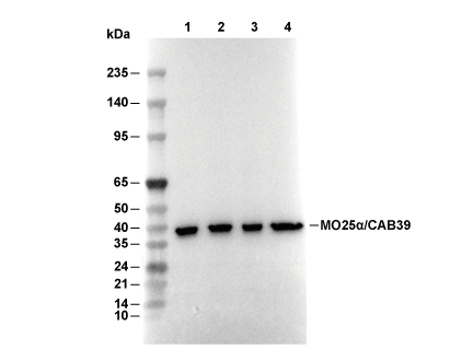 Lane 1: MCF-7, Lane 2: HepG2, Lane 3: C6, Lane 4: COS-7
Experiment Essentials
WB
Recommended wet transfer conditions: 200 mA, 60 min.
Recommended wet transfer conditions: 200 mA, 60 min.
Usage Information
| Dilution |
|---|
|
| Application |
|---|
| WB, IP |
| Reactivity |
|---|
| Human, Mouse, Rat, Monkey |
| Source |
|---|
| Rabbit Monoclonal Antibody |
| Storage Buffer |
|---|
| PBS, pH 7.2+50% Glycerol+0.05% BSA+0.01% NaN3 |
| Storage (from the date of receipt) |
|---|
| -20°C (avoid freeze-thaw cycles), 2 years |
| Predicted MW |
|---|
| 39 kDa |
| Positive Control | MCF-7 cells; HepG2 cells; HeLa cells; A204 cells; C6 cells; COS7 cells; L929 cells; C2C12 cells |
|---|---|
| Negative Control |
Experimental Methods
| WB |
|---|
Experimental Protocol:
Sample preparation
1. Tissue: Lyse the tissue sample by adding an appropriate volume of ice-cold RIPA/Tris-HCL Lysis Buffer (containing Protease Inhibitor Cocktail),and homogenize the tissue at a low temperature or lyse it by sonication on ice, then incubate on ice for 30 minutes. 2. Adherent cell: Aspirate the culture medium and wash the cells with ice-cold PBS twice. Lyse the cells by adding an appropriate volume of RIPA/Tris-HCL Lysis Buffer (containing Protease Inhibitor Cocktail) , sonicate to lyse the cells, and incubate on ice for 30 minutes. 3. Suspension cell: Transfer the culture medium to a pre-cooled centrifuge tube. Centrifuge and aspirate the supernatant. Wash the cells with ice-cold PBS twice. Lyse the cells by adding an appropriate volume of RIPA/Tris-HCL Lysis Buffer (containing Protease Inhibitor Cocktail) , sonicate to lyse the cells, and incubate on ice for 30 minutes. 4. Place the lysate into a pre-cooled microcentrifuge tube. Centrifuge at 4°C for 15 min. Collect the supernatant;
5. Remove a small volume of lysate to determine the protein concentration;
6. Combine the lysate with protein loading buffer. Boil 20 µL sample under 95-100°C for 5 min. Centrifuge for 5 min after cool down on ice.
Electrophoretic separation
1. According to the concentration of extracted protein, load appropriate amount of protein sample and marker onto SDS-PAGE gels for electrophoresis. Recommended separating gel (lower gel) concentration: 10%. Reference Table for Selecting SDS-PAGE Separation Gel Concentrations 2. Power up 80V for 30 minutes. Then the power supply is adjusted (110 V~150 V), the Marker is observed, and the electrophoresis can be stopped when the indicator band of the predyed protein Marker where the protein is located is properly separated. (Note that the current should not be too large when electrophoresis, too large current (more than 150 mA) will cause the temperature to rise, affecting the result of running glue. If high currents cannot be avoided, an ice bath can be used to cool the bath.)
Transfer membrane
1. Take out the converter, soak the clip and consumables in the pre-cooled converter;
2. Activate PVDF membrane with methanol for 1 min and rinse with transfer buffer;
3. Install it in the order of "black edge of clip - sponge - filter paper - filter paper - glue -PVDF membrane - filter paper - filter paper - sponge - white edge of clip"; 4. The protein was electrotransferred to PVDF membrane. ( 0.45 µm PVDF membrane is recommended ) Reference Table for Selecting PVDF Membrane Pore Size Specifications Recommended conditions for wet transfer: 200 mA, 60 min. ( Note that the transfer conditions can be adjusted according to the protein size. For high-molecular-weight proteins, a higher current and longer transfer time are recommended. However, ensure that the transfer tank remains at a low temperature to prevent gel melting.)
Block
1. After electrotransfer, wash the film with TBST at room temperature for 5 minutes;
2. Incubate the film in the blocking solution for 1 hour at room temperature;
3. Wash the film with TBST for 3 times, 5 minutes each time.
Antibody incubation
1. Use 5% skim milk powder to prepare the primary antibody working liquid (recommended dilution ratio for primary antibody 1:1000), gently shake and incubate with the film at 4°C overnight; 2. Wash the film with TBST 3 times, 5 minutes each time;
3. Add the secondary antibody to the blocking solution and incubate with the film gently at room temperature for 1 hour;
4. After incubation, wash the film with TBST 3 times for 5 minutes each time.
Antibody staining
1. Add the prepared ECL luminescent substrate (or select other color developing substrate according to the second antibody) and mix evenly;
2. Incubate with the film for 1 minute, remove excess substrate (keep the film moist), wrap with plastic film, and expose in the imaging system. |
Biological Description
| Specificity |
|---|
| CAB39/MO25 Antibody (Rabbit mAb) [H1E9] detects endogenous levels of total CAB39/MO25 protein. |
| Subcellular Location |
|---|
| Cytoplasm |
| Uniprot ID |
|---|
| Q9Y376 |
| Clone |
|---|
| H1E9 |
| Synonym(s) |
|---|
| Calcium-binding protein 39, MO25alpha, Protein Mo25, CAB39, MO25 |
| Background |
|---|
| CAB39, also known as MO25, is an armadillo repeat–containing scaffolding protein that forms a core component of the LKB1–STRAD–MO25 complex, which acts as the principal upstream kinase module for AMPK and a family of related kinases that regulate cellular energy sensing, polarity, and stress responses. The protein is built from tandem armadillo repeats that generate a curved surface for high‑affinity interaction with the C‑terminal Trp–Glu–Phe motif of the pseudokinase STRADα, and this binding markedly enhances the association of STRADα with the tumor suppressor kinase LKB1 and stabilizes the heterotrimeric complex. MO25 binding promotes proper folding and cytoplasmic localization of the STRAD–LKB1 complex and increases LKB1 catalytic activity toward downstream substrates, leading to phosphorylation and activation of AMPK and at least a dozen other AMPK‑related kinases that collectively control energy homeostasis, cell polarity, and metabolic adaptation under stress. LKB1–STRAD–MO25 is the major AMPK kinase assembly in tissues such as skeletal muscle, and MO25 acts as an essential cofactor that tunes both the stability and signaling competence of this node rather than contributing catalytic activity itself. CAB39/MO25 is also an important regulatory target for microRNAs that modulate the LKB1–AMPK axis: miR‑451 directly binds a conserved site in the CAB39 3′UTR, reduces CAB39 protein levels, and thereby diminishes LKB1‑dependent AMPK activation, shifting cells toward mTOR signaling and enhanced proliferation and survival in stress conditions. In cardiac and hypertrophic disease models, related microRNAs such as miR‑195 and miR‑451 target CAB39 transcripts and associate with reduced MO25 expression, lowered AMPK phosphorylation, and altered acetyl‑CoA carboxylase phosphorylation, linking CAB39 abundance to metabolic remodeling in the myocardium. |
| References |
|---|
|
Tech Support
Tel: +1-832-582-8158 Ext:3
If you have any other enquiries, please leave a message.

Products are for research use only. Not for human use. We do not sell to patients.
©Copyright 2013 Selleck Chemicals. All Rights Reserved.