-
 Australia
-
 Austria
-
 Belgium
-
 Brazil
-
 Canada
-
 China
China
-
 Czech Republic
-
 Denmark
-
 Finland
-
 France
-
 Germany
-
 Greece
-
 Hong Kong
-
 Hungary
-
 Iceland
-
 India
India
-
 Ireland
-
 Israel
-
 Italy
Italy
-
 Japan
Japan
-
 Korea
Korea
-
 Luxembourg
-
 Malaysia
-
 Netherlands
-
 New Zealand
-
 Norway
-
 Poland
-
 Qatar
Qatar
-
 Romania
-
 Saudi Arabia
-
 Singapore
-
 Spain
Spain
-
 Sweden
-
 Switzerland
-
 Taiwan
-
 Turkey
-
 United Kingdom
-
 United States
research use only
c-Rel Antibody [B22C17]
Cat.No.: F5039
Application:
Reactivity:
-
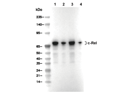 Lane 1: Raji, Lane 2: U973, Lane 3: A20, Lane 4: Neuro-2a
Usage Information
| Dilution |
|---|
|
| Application |
|---|
| WB |
| Reactivity |
|---|
| Human, Mouse, Rat |
| Source |
|---|
| Rabbit Monoclonal Antibody |
| Storage Buffer |
|---|
| PBS, pH 7.2+50% Glycerol+0.05% BSA+0.01% NaN3 |
| Storage (from the date of receipt) |
|---|
| -20°C (avoid freeze-thaw cycles), 2 years |
| Predicted MW |
|---|
| 68-78 kDa |
| Positive Control | Rat thymus; CCRF‑CEM cells; Raji cells; NK‑92 cells; U‑937 cells; A20 cells; Neuro‑2a cells. |
|---|---|
| Negative Control |
Experimental Methods
| WB |
|---|
Experimental Protocol:
Sample preparation
1. Tissue: Lyse the tissue sample by adding an appropriate volume of ice-cold RIPA/Nuclear Lysis Buffer (containing Protease Inhibitor Cocktail),and homogenize the tissue at a low temperature. 2. Adherent cell: Aspirate the culture medium and wash the cells with ice-cold PBS twice. Lyse the cells by adding an appropriate volume of RIPA/Nuclear Lysis Buffer (containing Protease Inhibitor Cocktail) and put the sample on ice for 5 min. 3. Suspension cell: Transfer the culture medium to a pre-cooled centrifuge tube. Centrifuge and aspirate the supernatant. Wash the cells with ice-cold PBS twice. Lyse the cells by adding an appropriate volume of RIPA/Nuclear Lysis Buffer (containing Protease Inhibitor Cocktail) and put the sample on ice for 5 min. 4. Place the lysate into a pre-cooled microcentrifuge tube. Centrifuge at 4°C for 15 min. Collect the supernatant;
5. Remove a small volume of lysate to determine the protein concentration;
6. Combine the lysate with protein loading buffer. Boil 20 µL sample under 95-100°C for 5 min. Centrifuge for 5 min after cool down on ice.
Electrophoretic separation
1. According to the concentration of extracted protein, load appropriate amount of protein sample and marker onto SDS-PAGE gels for electrophoresis. Recommended separating gel (lower gel) concentration: 10%. Reference Table for Selecting SDS-PAGE Separation Gel Concentrations 2. Power up 80V for 30 minutes. Then the power supply is adjusted (110 V~150 V), the Marker is observed, and the electrophoresis can be stopped when the indicator band of the predyed protein Marker where the protein is located is properly separated. (Note that the current should not be too large when electrophoresis, too large current (more than 150 mA) will cause the temperature to rise, affecting the result of running glue. If high currents cannot be avoided, an ice bath can be used to cool the bath.)
Transfer membrane
1. Take out the converter, soak the clip and consumables in the pre-cooled converter;
2. Activate PVDF membrane with methanol for 1 min and rinse with transfer buffer;
3. Install it in the order of "black edge of clip - sponge - filter paper - filter paper - glue -PVDF membrane - filter paper - filter paper - sponge - white edge of clip"; 4. The protein was electrotransferred to PVDF membrane. ( 0.45 µm PVDF membrane is recommended ) Reference Table for Selecting PVDF Membrane Pore Size Specifications Recommended conditions for wet transfer: 200 mA, 120 min. ( Note that the transfer conditions can be adjusted according to the protein size. For high-molecular-weight proteins, a higher current and longer transfer time are recommended. However, ensure that the transfer tank remains at a low temperature to prevent gel melting.)
Block
1. After electrotransfer, wash the film with TBST at room temperature for 5 minutes;
2. Incubate the film in the blocking solution for 1 hour at room temperature;
3. Wash the film with TBST for 3 times, 5 minutes each time.
Antibody incubation
1. Use 5% skim milk powder to prepare the primary antibody working liquid (recommended dilution ratio for primary antibody 1:1000), gently shake and incubate with the film at 4°C overnight; 2. Wash the film with TBST 3 times, 5 minutes each time;
3. Add the secondary antibody to the blocking solution and incubate with the film gently at room temperature for 1 hour;
4. After incubation, wash the film with TBST 3 times for 5 minutes each time.
Antibody staining
1. Add the prepared ECL luminescent substrate (or select other color developing substrate according to the second antibody) and mix evenly;
2. Incubate with the film for 1 minute, remove excess substrate (keep the film moist), wrap with plastic film, and expose in the imaging system. |
Biological Description
| Specificity |
|---|
| c-Rel Antibody [B22C17] detects endogenous levels of total c-Rel protein. |
| Subcellular Location |
|---|
| Nucleus |
| Uniprot ID |
|---|
| Q04864 |
| Clone |
|---|
| B22C17 |
| Synonym(s) |
|---|
| Proto-oncogene c-Rel; REL |
| Background |
|---|
| c-Rel belongs to the Rel/NF-κB family of transcription factors characterized by a Rel homology domain that mediates DNA binding, dimerization, and nuclear localization. The protein contains an N-terminal Rel homology domain encompassing DNA-binding and dimerization regions, along with C-terminal transactivation domains unique to RelA, c-Rel, and RelB members. In resting hematopoietic cells where c-Rel expression predominates, IκBα and IκBβ sequester c-Rel dimers in the cytoplasm through binding to the nuclear localization signal within the Rel homology domain. Proinflammatory stimuli trigger the IKK complex to phosphorylate IκBα at N-terminal serines, leading to its K48-linked ubiquitination and proteasomal degradation, which unmasks the c-Rel nuclear localization signal and enables nuclear translocation as p50/c-Rel or RelA/c-Rel heterodimers. These dimers bind κB sites in promoters of target genes encoding cytokines, anti-apoptotic factors, and cell cycle regulators via the Rel homology domain's DNA-binding region. Distinct domains of IκBα mediate cytoplasmic retention through N-terminal and central ankyrin repeats that mask the nuclear localization signal, while central ankyrin and C-terminal acidic residues suffice for nuclear inhibition by competing for DNA-binding sites or corepressors. c-Rel forms stable p65/c-Rel heterodimers with IκBβ in unstimulated states, where hyperphosphorylated IκBβ acts as an inhibitor until stimulus-induced slow phosphorylation triggers its partial degradation, selectively releasing p65/c-Rel for TNFα promoter activation. Nuclear IκBα colocalizes with constitutively nuclear c-Rel variants to suppress transactivation. In T cell differentiation, c-Rel induces RORγt and IL-21 expression critical for Th17 and Tfh lineages, while supporting Foxp3 in regulatory T cells. Elevated c-Rel activity drives lymphoid malignancies and chronic inflammation through persistent target gene expression. |
| References |
|---|
|
Tech Support
Tel: +1-832-582-8158 Ext:3
If you have any other enquiries, please leave a message.
Products are for research use only. Not for human use. We do not sell to patients.
©Copyright 2013 Selleck Chemicals. All Rights Reserved.