
-
 Australia
-
 Austria
-
 Belgium
-
 Brazil
-
 Canada
-
 China
China
-
 Czech Republic
-
 Denmark
-
 Finland
-
 France
-
 Germany
-
 Greece
-
 Hong Kong
-
 Hungary
-
 Iceland
-
 India
India
-
 Ireland
-
 Israel
-
 Italy
Italy
-
 Japan
Japan
-
 Korea
Korea
-
 Luxembourg
-
 Malaysia
-
 Netherlands
-
 New Zealand
-
 Norway
-
 Poland
-
 Qatar
Qatar
-
 Romania
-
 Saudi Arabia
-
 Singapore
-
 Spain
Spain
-
 Sweden
-
 Switzerland
-
 Taiwan
-
 Turkey
-
 United Kingdom
-
 United States
research use only
C-Peptide Antibody (Rabbit mAb) [H2G21]
CatNo: F5835
Application:
Reactivity:
-
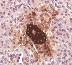 Immunohistochemical analysis of formalin fixed paraffin embedded human pancreas tissue with F5835 at 1:10000 dilution.
Usage Information
| Dilution |
|---|
|
| Application |
|---|
| IHC, ELISA |
| Reactivity |
|---|
| Mouse, Rat, Human |
| Source |
|---|
| Rabbit Monoclonal Antibody |
| Storage Buffer |
|---|
| PBS, pH 7.2+50% Glycerol+0.05% BSA+0.01% NaN3 |
| Storage (from the date of receipt) |
|---|
| -20°C (avoid freeze-thaw cycles), 2 years |
| Predicted MW |
|---|
| 12 kDa |
| Positive Control | Mouse pancreas; Human pancreas tissue; Rat pancreas tissue; Recombinant mature C-Peptide protein |
|---|---|
| Negative Control |
Experimental Methods
| IHC |
|---|
Experimental Protocol:
Deparaffinization/Rehydration
1. Deparaffinize/hydrate sections:
2. Incubate sections in three washes of xylene for 5 min each.
3. Incubate sections in two washes of 100% ethanol for 10 min each.
4. Incubate sections in two washes of 95% ethanol for 10 min each.
5. Wash sections two times in dH2O for 5 min each.
6.Antigen retrieval: For Citrate: Heat slides in a microwave submersed in 1X citrate unmasking solution until boiling is initiated; continue with 10 min at a sub-boiling temperature (95°-98°C). Cool slides on bench top for 30 min.
Staining
1. Wash sections in dH2O three times for 5 min each.
2. Incubate sections in 3% hydrogen peroxide for 10 min.
3. Wash sections in dH2O two times for 5 min each.
4. Wash sections in wash buffer for 5 min.
5. Block each section with 100–400 µl of blocking solution for 1 hr at room temperature.
6. Remove blocking solution and add 100–400 µl primary antibody diluent in to each section. Incubate overnight at 4°C.
7. Remove antibody solution and wash sections with wash buffer three times for 5 min each.
8. Cover section with 1–3 drops HRPas needed. Incubate in a humidified chamber for 30 min at room temperature.
9. Wash sections three times with wash buffer for 5 min each.
10. Add DAB Chromogen Concentrate to DAB Diluent and mix well before use.
11. Apply 100–400 µl DAB to each section and monitor closely. 1–10 min generally provides an acceptable staining intensity.
12. Immerse slides in dH2O.
13. If desired, counterstain sections with hematoxylin.
14. Wash sections in dH2O two times for 5 min each.
15. Dehydrate sections: Incubate sections in 95% ethanol two times for 10 sec each; Repeat in 100% ethanol, incubating sections two times for 10 sec each; Repeat in xylene, incubating sections two times for 10 sec each.
16. Mount sections with coverslips and mounting medium.
|
Biological Description
| Specificity |
|---|
| C-Peptide Antibody (Rabbit mAb) [H2G21] detects endogenous C-peptide, an insulin-processing peptide. |
| Subcellular Location |
|---|
| Secreted |
| Uniprot ID |
|---|
| P01308 |
| Clone |
|---|
| H2G21 |
| Synonym(s) |
|---|
| Insulin |
| Background |
|---|
| Proinsulin C-peptide is the connecting segment excised during proinsulin processing and released from pancreatic beta cells in equimolar amounts with insulin, placing it structurally within the insulin family while distinguishing it functionally as an independent signaling entity rather than an inert processing remnant. C-peptide binds cell surfaces in the nanomolar range through a stereospecific interaction that shows no cross-reactivity with insulin, proinsulin, or related growth factors, consistent with engagement of a distinct, most likely G-protein-coupled receptor rather than the canonical insulin receptor. Receptor engagement is pertussis-toxin sensitive, pointing to coupling through Gαi/o, yet C-peptide also activates protein kinase C, a pathway ordinarily linked to Gαq-coupled receptors, and this pattern is explained by G-protein promiscuity in which the receptor shifts coupling preference according to the relative extracellular concentrations of insulin and C-peptide. In settings of low insulin, receptor coupling favors Gαi/o and produces enhanced activation of insulin-related MAPK signaling together with downstream Akt activity, whereas in settings of high insulin the receptor favors Gαq coupling, driving PKC activation that subsequently dampens insulin signaling through serine/threonine phosphorylation of IRS-1, positioning C-peptide as a concentration-dependent modulator that fine-tunes insulin signal strength rather than acting as a static bystander. Receptor binding further triggers a rise in intracellular calcium, increased phosphoinositide-3-kinase activity, stimulation of Na+/K+-ATPase, induction of endothelial nitric oxide synthase transcription, and engagement of the MAPK cascade, and these downstream effects converge functionally on vasodilation, anti-inflammatory action, anti-apoptotic protection, and antioxidant activity within the vascular endothelium, with additional influence extending to erythrocytes and immune cells through both direct and indirect receptor-mediated routes. The orphan receptor GPR146 has been identified as a component of this C-peptide signalosome, since its knockdown abolishes C-peptide-induced immediate-early gene expression and receptor-directed antibodies block C-peptide-specific downstream effects without altering insulin action, marking GPR146 as a candidate mediator of C-peptide's tissue-specific signaling. Diabetic complications, including peripheral nerve and renal dysfunction, correlate with reduced Na+/K+-ATPase activity, and restoring C-peptide in insulin-deficient states reverses these functional and structural abnormalities, linking the receptor-mediated signaling cascade directly to the peptide's protective actions in the kidney, nerve, and microvasculature and underscoring its relevance as a target for evaluating tissue-specific insulin-adjacent signaling. |
| References |
|---|
|
Tech Support
Tel: +1-832-582-8158 Ext:3
If you have any other enquiries, please leave a message.

Products are for research use only. Not for human use. We do not sell to patients.
©Copyright 2013 Selleck Chemicals. All Rights Reserved.