
-
 Argentina
-
 Australia
-
 Austria
-
 Belgium
-
 Brazil
-
 Canada
-
 China
China
-
 Czech Republic
-
 Denmark
-
 Finland
-
 France
-
 Germany
-
 Greece
-
 Hong Kong
-
 Hungary
-
 Iceland
-
 India
India
-
 Ireland
-
 Israel
-
 Italy
Italy
-
 Japan
Japan
-
 Korea
Korea
-
 Luxembourg
-
 Malaysia
-
 Netherlands
-
 New Zealand
-
 Norway
-
 Poland
-
 Portugal
-
 Qatar
Qatar
-
 Romania
-
 Saudi Arabia
-
 Singapore
-
 Spain
Spain
-
 Sweden
-
 Switzerland
-
 Taiwan
-
 Turkey
-
 United Kingdom
-
 United States
research use only
β Amyloid (Aβ) 1-42 Antibody (Rabbit mAb) [M4F1]
CatNo: F1146
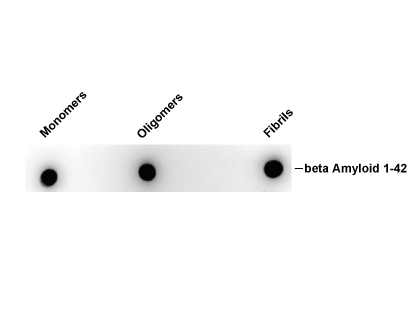
Usage Information
| Dilution |
|---|
|
| Application |
|---|
| IHC, Dot |
| Reactivity |
|---|
| Mouse, Human |
| Source |
|---|
| Rabbit Monoclonal Antibody |
| Storage Buffer |
|---|
| PBS, pH 7.2+50% Glycerol+0.05% BSA+0.01% NaN3 |
| Storage (from the date of receipt) |
|---|
| -20°C (avoid freeze-thaw cycles), 2 years |
| Predicted MW |
|---|
| 87 kDa |
| Positive Control | Human Alzheimer hippocampus |
|---|---|
| Negative Control |
Experimental Methods
| IHC |
|---|
Experimental Protocol:
Deparaffinization/Rehydration
1. Deparaffinize/hydrate sections:
2. Incubate sections in three washes of xylene for 5 min each.
3. Incubate sections in two washes of 100% ethanol for 10 min each.
4. Incubate sections in two washes of 95% ethanol for 10 min each.
5. Wash sections two times in dH2O for 5 min each.
6.Antigen retrieval: For Citrate: Heat slides in a microwave submersed in 1X citrate unmasking solution until boiling is initiated; continue with 10 min at a sub-boiling temperature (95°-98°C). Cool slides on bench top for 30 min.
Staining
1. Wash sections in dH2O three times for 5 min each.
2. Incubate sections in 3% hydrogen peroxide for 10 min.
3. Wash sections in dH2O two times for 5 min each.
4. Wash sections in wash buffer for 5 min.
5. Block each section with 100–400 µl of blocking solution for 1 hr at room temperature.
6. Remove blocking solution and add 100–400 µl primary antibody diluent in to each section. Incubate overnight at 4°C.
7. Remove antibody solution and wash sections with wash buffer three times for 5 min each.
8. Cover section with 1–3 drops HRPas needed. Incubate in a humidified chamber for 30 min at room temperature.
9. Wash sections three times with wash buffer for 5 min each.
10. Add DAB Chromogen Concentrate to DAB Diluent and mix well before use.
11. Apply 100–400 µl DAB to each section and monitor closely. 1–10 min generally provides an acceptable staining intensity.
12. Immerse slides in dH2O.
13. If desired, counterstain sections with hematoxylin.
14. Wash sections in dH2O two times for 5 min each.
15. Dehydrate sections: Incubate sections in 95% ethanol two times for 10 sec each; Repeat in 100% ethanol, incubating sections two times for 10 sec each; Repeat in xylene, incubating sections two times for 10 sec each.
16. Mount sections with coverslips and mounting medium.
|
Biological Description
| Specificity |
|---|
β Amyloid (Aβ) 1-42 Antibody (Rabbit mAb) [M4F1] detects endogenous levels of total β Amyloid (Aβ) 1-42 protein. |
| Subcellular Location |
|---|
| Amyloid, Cell membrane, Cell projection, Coated pit, Cytoplasm, Endoplasmic reticulum, Endosome, Golgi apparatus |
| Uniprot ID |
|---|
| P05067 |
| Clone |
|---|
| M4F1 |
| Synonym(s) |
|---|
| A4, AD1, APP, Amyloid-beta precursor protein, ABPP, APPI, Alzheimer disease amyloid A4 protein homolog, Cerebral vascular amyloid peptide, PreA4, Protease nexin-II, CVAP, PN-II |
| Background |
|---|
β-Amyloid 1-42 is a peptide fragment released from the amyloid precursor protein through sequential proteolytic cleavage, representing the primary product of the amyloidogenic processing branch of APP metabolism. Full-length APP undergoes cleavage by β-secretase (BACE1) at the cell surface and within endocytic compartments, generating a soluble N-terminal fragment, sAPPβ, and a membrane-retained C-terminal fragment, C99. Sequential cleavage of C99 by the γ-secretase complex, composed of presenilin, nicastrin, Pen-2, and Aph-1 subunits, releases Aβ into the extracellular space along with the intracellular domain AICD, which itself acts as a transcriptional regulator influencing further APP expression. This amyloidogenic route operates in competition with the non-amyloidogenic pathway, where α-secretase cleavage within the Aβ domain precludes intact peptide formation and instead yields the neuroprotective fragment sAPPα, positioning secretase balance as the determinant of Aβ output. Once generated, Aβ1-42 self-associates into soluble oligomers and higher-order fibrils that accumulate as extracellular plaques, interacting with neuronal membrane lipids, ion channels, and surface receptors to disrupt synaptic signaling and calcium homeostasis. Aβ pathology intersects with tau biology through shared kinase signaling intermediates that promote tau hyperphosphorylation, driving microtubule destabilization and neurofibrillary tangle formation as a parallel pathological cascade. BACE1 cleavage constitutes the rate-limiting step of the amyloidogenic pathway, and its activity is influenced by cellular trafficking, membrane lipid raft composition, and stress-responsive kinase signaling, including JNK and p38 MAPK pathways that elevate BACE1 expression under oxidative conditions. Aβ1-42 also accumulates within cerebral blood vessel walls, contributing to cerebral amyloid angiopathy as a distinct but overlapping vascular pathology. |
| References |
|---|
|
Tech Support
Tel: +1-832-582-8158 Ext:3
If you have any other enquiries, please leave a message.

Products are for research use only. Not for human use. We do not sell to patients.
©Copyright 2013 Selleck Chemicals. All Rights Reserved.