-
 Australia
-
 Austria
-
 Belgium
-
 Brazil
-
 Canada
-
 China
China
-
 Czech Republic
-
 Denmark
-
 Finland
-
 France
-
 Germany
-
 Greece
-
 Hong Kong
-
 Hungary
-
 Iceland
-
 India
India
-
 Ireland
-
 Israel
-
 Italy
Italy
-
 Japan
Japan
-
 Korea
Korea
-
 Luxembourg
-
 Malaysia
-
 Netherlands
-
 New Zealand
-
 Norway
-
 Poland
-
 Qatar
Qatar
-
 Romania
-
 Saudi Arabia
-
 Singapore
-
 Spain
Spain
-
 Sweden
-
 Switzerland
-
 Taiwan
-
 Turkey
-
 United Kingdom
-
 United States
research use only
Argonaute-2 Antibody [K4M16]
Cat.No.: F4172
Application:
Reactivity:
-
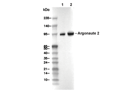 Lane 1: MCF-7, Lane 2: HepG2
Usage Information
| Dilution |
|---|
|
| Application |
|---|
| WB, IP |
| Reactivity |
|---|
| Human, Mouse, Rat, Monkey |
| Source |
|---|
| Rabbit Monoclonal Antibody |
| Storage Buffer |
|---|
| PBS, pH 7.2+50% Glycerol+0.05% BSA+0.01% NaN3 |
| Storage (from the date of receipt) |
|---|
| -20°C (avoid freeze-thaw cycles), 2 years |
| Predicted MW |
|---|
| 97 kDa |
| Positive Control | MCF-7 cells; HepG2 cells; mIMCD3 cells; KNRK cells; COS-7 cells |
|---|---|
| Negative Control |
Experimental Methods
| WB |
|---|
Experimental Protocol:
Sample preparation
1. Tissue: Lyse the tissue sample by adding an appropriate volume of ice-cold RIPA/NP-40 Lysis Buffer (containing Protease Inhibitor Cocktail),and homogenize the tissue at a low temperature or lyse it by sonication on ice, then incubate on ice for 30 minutes. 2. Adherent cell: Aspirate the culture medium and wash the cells with ice-cold PBS twice. Lyse the cells by adding an appropriate volume of RIPA/NP-40 Lysis Buffer (containing Protease Inhibitor Cocktail), sonicate to lyse the cells, and incubate on ice for 30 minutes. 3. Suspension cell: Transfer the culture medium to a pre-cooled centrifuge tube. Centrifuge and aspirate the supernatant. Wash the cells with ice-cold PBS twice. Lyse the cells by adding an appropriate volume of RIPA/NP-40 Lysis Buffer (containing Protease Inhibitor Cocktail), sonicate to lyse the cells, and incubate on ice for 30 minutes. 4. Place the lysate into a pre-cooled microcentrifuge tube. Centrifuge at 4°C for 15 min. Collect the supernatant;
5. Remove a small volume of lysate to determine the protein concentration;
6. Combine the lysate with protein loading buffer. Boil 20 µL sample under 95-100°C for 5 min. Centrifuge for 5 min after cool down on ice.
Electrophoretic separation
1. According to the concentration of extracted protein, load appropriate amount of protein sample and marker onto SDS-PAGE gels for electrophoresis. Recommended separating gel (lower gel) concentration: 10%. Reference Table for Selecting SDS-PAGE Separation Gel Concentrations 2. Power up 80V for 30 minutes. Then the power supply is adjusted (110 V~150 V), the Marker is observed, and the electrophoresis can be stopped when the indicator band of the predyed protein Marker where the protein is located is properly separated. (Note that the current should not be too large when electrophoresis, too large current (more than 150 mA) will cause the temperature to rise, affecting the result of running glue. If high currents cannot be avoided, an ice bath can be used to cool the bath.)
Transfer membrane
1. Take out the converter, soak the clip and consumables in the pre-cooled converter;
2. Activate PVDF membrane with methanol for 1 min and rinse with transfer buffer;
3. Install it in the order of "black edge of clip - sponge - filter paper - filter paper - glue -PVDF membrane - filter paper - filter paper - sponge - white edge of clip"; 4. The protein was electrotransferred to PVDF membrane. ( 0.45 µm PVDF membrane is recommended ) Reference Table for Selecting PVDF Membrane Pore Size Specifications Recommended conditions for wet transfer: 200 mA, 120 min. ( Note that the transfer conditions can be adjusted according to the protein size. For high-molecular-weight proteins, a higher current and longer transfer time are recommended. However, ensure that the transfer tank remains at a low temperature to prevent gel melting.)
Block
1. After electrotransfer, wash the film with TBST at room temperature for 5 minutes;
2. Incubate the film in the blocking solution for 1 hour at room temperature;
3. Wash the film with TBST for 3 times, 5 minutes each time.
Antibody incubation
1. Use 5% skim milk powder to prepare the primary antibody working liquid (recommended dilution ratio for primary antibody 1:1000), gently shake and incubate with the film at 4°C overnight; 2. Wash the film with TBST 3 times, 5 minutes each time;
3. Add the secondary antibody to the blocking solution and incubate with the film gently at room temperature for 1 hour;
4. After incubation, wash the film with TBST 3 times for 5 minutes each time.
Antibody staining
1. Add the prepared ECL luminescent substrate (or select other color developing substrate according to the second antibody) and mix evenly;
2. Incubate with the film for 1 minute, remove excess substrate (keep the film moist), wrap with plastic film, and expose in the imaging system. |
Biological Description
| Specificity |
|---|
| Argonaute-2 Antibody [K4M16] detects endogenous levels of total Argonaute-2 protein. |
| Subcellular Location |
|---|
| Cytoplasm, Nucleus |
| Uniprot ID |
|---|
| Q8CJG0 |
| Clone |
|---|
| K4M16 |
| Synonym(s) |
|---|
| Protein argonaute-2; Argonaute 2; mAgo2; Argonaute RISC catalytic component 2; eIF-2C 2; eIF2C 2; Piwi/argonaute family protein meIF2C2; Protein slicer; Ago2; Eif2c2; Kiaa4215 |
| Background |
|---|
| Argonaute-2, or Ago2, is the only member of the mammalian Argonaute protein family (AGO1-4) with intrinsic endonuclease or slicer activity, making it the central effector of the RNA-induced silencing complex, or RISC, which mediates RNA interference pathways using small RNA guides such as microRNAs, small interfering RNAs, and piwi-interacting RNAs for gene silencing. Ago2 adopts a bi-lobed architecture composed of four core domains: the N domain, which is involved in guide strand unwinding; the PAZ domain, which anchors the 3' end of the guide RNA; the MID domain, which anchors the 5' end; and the PIWI domain, which is structurally similar to RNase H and houses the catalytic activity. These domains are connected by L1 and L2 linker regions. The PIWI domain contains a catalytic triad of aspartate, glutamate, and aspartate residues that coordinate two magnesium ions necessary for mRNA cleavage between guide positions 10 and 11, while specific residues such as arginine 635 and glutamine 633 in the seed-pairing region ensure target specificity by introducing kinks in the RNA duplex. Ago2 loads mature double-stranded miRNA or siRNA in the cytoplasm, selects the guide strand based on thermodynamic stability, and threads it through a central cleft where it interacts with all domains and linkers for enhanced stability. Depending on the degree of complementarity, Ago2 can execute endonucleolytic slicing for perfectly matched targets or mediate translational repression and deadenylation for mismatched targets via recruitment of GW182. Multiple phosphorylation sites modulate Ago2's localization, RNA-binding affinity, and silencing efficiency. Ago2 is essential for development, differentiation, cellular stress responses, and genome stability by silencing a substantial fraction of human genes. It orchestrates RISC assembly with Dicer and TRBP and also plays roles in chromatin silencing. Dysregulation of Ago2 can promote oncogenesis, as overexpression is seen in several cancers and enhances cell proliferation and invasion by stabilizing oncogenic microRNAs like miR-21 and miR-155, while it is also implicated in neurodegenerative diseases. |
| References |
|---|
|
Tech Support
Tel: +1-832-582-8158 Ext:3
If you have any other enquiries, please leave a message.
Products are for research use only. Not for human use. We do not sell to patients.
©Copyright 2013 Selleck Chemicals. All Rights Reserved.