-
 Australia
-
 Austria
-
 Belgium
-
 Brazil
-
 Canada
-
 China
China
-
 Czech Republic
-
 Denmark
-
 Finland
-
 France
-
 Germany
-
 Greece
-
 Hong Kong
-
 Hungary
-
 Iceland
-
 India
India
-
 Ireland
-
 Israel
-
 Italy
Italy
-
 Japan
Japan
-
 Korea
Korea
-
 Luxembourg
-
 Malaysia
-
 Netherlands
-
 New Zealand
-
 Norway
-
 Poland
-
 Qatar
Qatar
-
 Romania
-
 Saudi Arabia
-
 Singapore
-
 Spain
Spain
-
 Sweden
-
 Switzerland
-
 Taiwan
-
 Turkey
-
 United Kingdom
-
 United States
research use only
ADAM9 Antibody [M15P12]
Cat.No.: F9371
Application:
Reactivity:
-
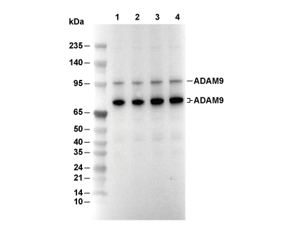 Lane 1: Hela, Lane 2: PC12, Lane 3: NIH/3T3, Lane 4: C2C12
Usage Information
| Dilution |
|---|
|
| Application |
|---|
| WB |
| Reactivity |
|---|
| Human, Mouse, Rat, Monkey |
| Source |
|---|
| Rabbit Monoclonal Antibody |
| Storage Buffer |
|---|
| PBS, pH 7.2+50% Glycerol+0.05% BSA+0.01% NaN3 |
| Storage (from the date of receipt) |
|---|
| -20°C (avoid freeze-thaw cycles), 2 years |
| Predicted MW |
|---|
| 100-115 kDa, 75-80 kDa |
| Positive Control | HeLa cells; COS-7 cells; C6 cells; PC12 cells; MTLN3 cells; NIH/3T3 cells; C2C12 cells |
|---|---|
| Negative Control |
Experimental Methods
| WB |
|---|
Experimental Protocol:
Sample preparation
1. Tissue: Lyse the tissue sample by adding an appropriate volume of ice-cold RIPA/NP-40 Lysis Buffer (containing Protease Inhibitor Cocktail),and homogenize the tissue at a low temperature. 2. Adherent cell: Aspirate the culture medium and wash the cells with ice-cold PBS twice. Lyse the cells by adding an appropriate volume of RIPA/NP-40 Lysis Buffer (containing Protease Inhibitor Cocktail) and put the sample on ice for 5 min. 3. Suspension cell: Transfer the culture medium to a pre-cooled centrifuge tube. Centrifuge and aspirate the supernatant. Wash the cells with ice-cold PBS twice. Lyse the cells by adding an appropriate volume of RIPA/NP-40 Lysis Buffer (containing Protease Inhibitor Cocktail) and put the sample on ice for 5 min. 4. Place the lysate into a pre-cooled microcentrifuge tube. Centrifuge at 4°C for 15 min. Collect the supernatant;
5. Remove a small volume of lysate to determine the protein concentration;
6. Combine the lysate with protein loading buffer. Boil 20 µL sample under 95-100°C for 5 min. Centrifuge for 5 min after cool down on ice.
Electrophoretic separation
1. According to the concentration of extracted protein, load appropriate amount of protein sample and marker onto SDS-PAGE gels for electrophoresis. Recommended separating gel (lower gel) concentration: 10%. Reference Table for Selecting SDS-PAGE Separation Gel Concentrations 2. Power up 80V for 30 minutes. Then the power supply is adjusted (110 V~150 V), the Marker is observed, and the electrophoresis can be stopped when the indicator band of the predyed protein Marker where the protein is located is properly separated. (Note that the current should not be too large when electrophoresis, too large current (more than 150 mA) will cause the temperature to rise, affecting the result of running glue. If high currents cannot be avoided, an ice bath can be used to cool the bath.)
Transfer membrane
1. Take out the converter, soak the clip and consumables in the pre-cooled converter;
2. Activate PVDF membrane with methanol for 1 min and rinse with transfer buffer;
3. Install it in the order of "black edge of clip - sponge - filter paper - filter paper - glue -PVDF membrane - filter paper - filter paper - sponge - white edge of clip"; 4. The protein was electrotransferred to PVDF membrane. ( 0.45 µm PVDF membrane is recommended ) Reference Table for Selecting PVDF Membrane Pore Size Specifications Recommended conditions for wet transfer: 200 mA, 120 min. ( Note that the transfer conditions can be adjusted according to the protein size. For high-molecular-weight proteins, a higher current and longer transfer time are recommended. However, ensure that the transfer tank remains at a low temperature to prevent gel melting.)
Block
1. After electrotransfer, wash the film with TBST at room temperature for 5 minutes;
2. Incubate the film in the blocking solution for 1 hour at room temperature;
3. Wash the film with TBST for 3 times, 5 minutes each time.
Antibody incubation
1. Use 5% skim milk powder to prepare the primary antibody working liquid (recommended dilution ratio for primary antibody 1:1000), gently shake and incubate with the film at 4°C overnight; 2. Wash the film with TBST 3 times, 5 minutes each time;
3. Add the secondary antibody to the blocking solution and incubate with the film gently at room temperature for 1 hour;
4. After incubation, wash the film with TBST 3 times for 5 minutes each time.
Antibody staining
1. Add the prepared ECL luminescent substrate (or select other color developing substrate according to the second antibody) and mix evenly;
2. Incubate with the film for 1 minute, remove excess substrate (keep the film moist), wrap with plastic film, and expose in the imaging system. |
Biological Description
| Specificity |
|---|
| ADAM9 Antibody [M15P12] detects endogenous levels of total ADAM9 protein. |
| Subcellular Location |
|---|
| Cell membrane, Membrane, Secreted |
| Uniprot ID |
|---|
| Q13443 |
| Clone |
|---|
| M15P12 |
| Synonym(s) |
|---|
| Disintegrin and metalloproteinase domain-containing protein 9; ADAM 9; Cellular disintegrin-related protein; Meltrin-gamma; Metalloprotease/disintegrin/cysteine-rich protein 9; Myeloma cell metalloproteinase; ADAM9; KIAA0021; MCMP; MDC9; MLTNG |
| Background |
|---|
| ADAM9, known as Meltrin gamma, belongs to the ADAM family of transmembrane metalloproteases that combine proteolytic and cell adhesion capabilities. The protein spans 819 amino acids with an 84 kDa mature mass, organized into a signal peptide (1–29), prodomain (30–205) that maintains latency via cysteine-switch, zinc-dependent metalloprotease domain (212–427) with HEXXHXXGXXH catalytic motif, disintegrin domain (413–503) featuring integrin-binding motifs, cysteine-rich domain (507–627), EGF-like repeats, transmembrane helix (707–728), and cytoplasmic tail with SH3-binding proline-rich region. Furin-like convertases cleave the prodomain at RSKRR125↓SV to activate proteolysis, while ADAM9 homodimerizes via cysteine-rich domains to expose the active site. ADAM9 sheds ectodomains of EGFR ligands (HB-EGF, TGF-α), MET, ErbB3/4, Notch, and APP, releasing soluble forms that drive proliferation, migration, and survival signaling through MAPK/ERK, PI3K/AKT, and Notch pathways. The disintegrin domain binds α6β1, αvβ3/6, and α2β1 integrins to modulate adhesion, migration, and invasion independent of catalysis. Cysteine-rich and EGF-like regions interact with syndecans and ECM components to regulate matrix remodeling during angiogenesis and wound healing. Cytoplasmic interactions with Src, PKC, and tetraspanins (CD9, CD151) fine-tune trafficking and activity at invadopodia. ADAM9 governs fertilization by cleaving ZP2/3, myogenesis via Notch/MEF2C, retinal pigment epithelium differentiation, and immune cell diapedesis. In cancer, upregulated ADAM9 in breast, prostate, lung, pancreas, and brain tumors promotes metastasis via EGFR activation, EMT, and angiogenesis; knockout reduces invasion by 80%. ADAM9 processes APP to neuroprotectin fragments, countering amyloid-β accumulation. Inflammatory roles emerge in rheumatoid arthritis via integrin shedding and macrophage infiltration. These actions position ADAM9 as a pivotal regulator of adhesion dynamics and growth factor bioavailability across development and pathology. |
| References |
|---|
|
Tech Support
Tel: +1-832-582-8158 Ext:3
If you have any other enquiries, please leave a message.
Products are for research use only. Not for human use. We do not sell to patients.
©Copyright 2013 Selleck Chemicals. All Rights Reserved.