|
How to Cite 1. For In-Text Citation (Materials & Methods): 2. For Key Resources Table: |
||
|
Toll Free: (877) 796-6397 -- USA and Canada only -- |
Fax: +1-832-582-8590 Orders: +1-832-582-8158 |
Tech Support: +1-832-582-8158 Ext:3 Please provide your Order Number in the email. We strive to reply to |
Technical Data
| Formula | C14H20N2O3 |
||||||
| Molecular Weight | 264.3 | CAS No. | 149647-78-9 | ||||
| Solubility (25°C)* | In vitro | DMSO | 52 mg/mL (196.74 mM) | ||||
| Ethanol | 6.5 mg/mL (24.59 mM) | ||||||
| Water | Insoluble | ||||||
| In vivo (Add solvents to the product individually and in order) |
|
||||||
|
* <1 mg/ml means slightly soluble or insoluble. * Please note that Selleck tests the solubility of all compounds in-house, and the actual solubility may differ slightly from published values. This is normal and is due to slight batch-to-batch variations. * Room temperature shipping (Stability testing shows this product can be shipped without any cooling measures.) |
|||||||
Preparing Stock Solutions
Biological Activity
| Description | Vorinostat (SAHA) is an HDAC inhibitor with IC50 of ~10 nM in a cell-free assay and abrogates productive HPV-18 DNA amplification. | ||
|---|---|---|---|
| Targets |
|
||
| In vitro | Vorinostat (SAHA) inhibits the activities of HDAC1 and HDAC3 with IC50 of 10 nM and 20 nM, respectively, and also results in a marked hyperacetylation of histone H4. This compound inhibits the growth of three prostate cancer cell lines LNCaP, PC-3 and TSU-Pr1 at micromolar concentrations (2.5-7.5 μM), and induces dose-dependent cell death in LNCaP cells. Its treatment in MCF-7 cells inhibits cell proliferation at an IC50 of 0.75 μM resulting in the accumulation of cells in the G1 and G2-M phase of the cell cycle. It also induces differentiation in the estrogen receptor-negative cell line SKBr-3 and the retinoblastoma-negative cell line MDA-468. Treatment with this compound at 1 μM for 8 hours or more is sufficient to irreversibly induce apoptosis of human multiple myeloma (MM) cells. The gene expression profiles of Vorinostat-treated MM cells are not hallmarked by global transcriptional activation, but by coordinated transcriptional changes of specific functional groups of genes such as cytokine-induced proliferative/survival signaling cascades, oncogenes-tumor suppressor genes, regulators of apoptosis, DNA synthesis-repair and cell cycle, and proteasome-ubiquitin function. |
||
| In vivo | Administration of Vorinostat (SAHA) (~100 mg/kg/day) significantly inhibits the growth of CWR22 human prostate xenografts in nude mice with tumor reductions of 78%, 97% and 97%, at doses of 25 mg/kg/day, 50 mg/kg/day and 100 mg/kg/day, respectively, compared with control. This compound induces the accumulation of acetylated core histones and prostate-specific antigen mRNA expression in CWR22 cells, resulting in higher levels of serum prostate-specific antigen than predicted from tumor volume alone. Oral administration of it (0.67g/L) crosses the blood-brain barrier, increases histone acetylation in the brain, and dramatically improves the motor impairment in the R6/2 mice model of Huntington's disease. |
Protocol (from reference)
| Kinase Assay: |
|
|---|---|
| Cell Assay: |
|
| Animal Study: |
|
References
|
Customer Product Validation
-
Data from [ J Exp Med , 2012 , 209, 35-50 ]
-
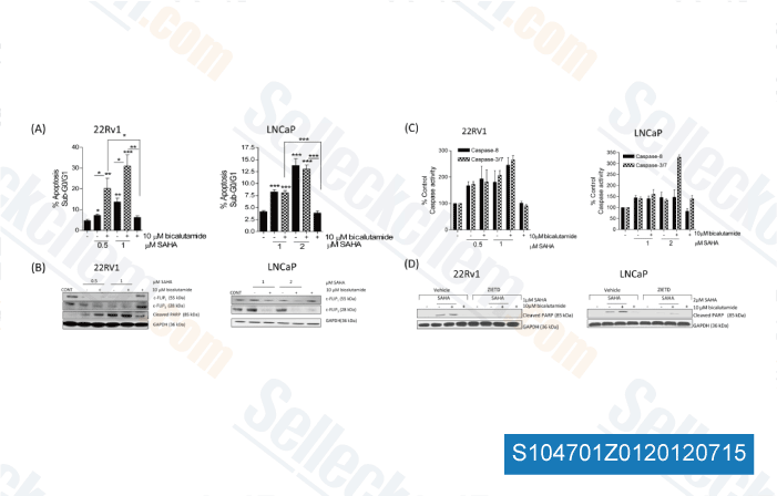
Data from [ Clin Cancer Res , 2012 , 18, 3822-33 ]
-
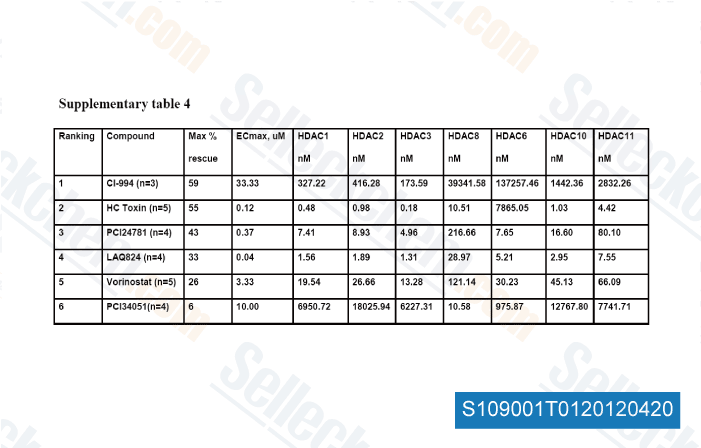
Data from [ Diabetologia , 2012 , 55, 2421-2431 ]
-
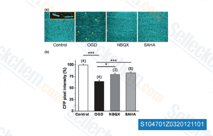
Data from [ J Neurochem , 2012 , 123 Suppl 2, 108-15 ]
Selleck's Vorinostat (SAHA) Has Been Cited by 618 Publications
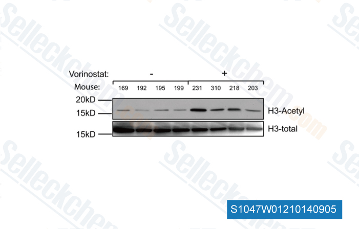
| Vorinostat attenuates UVB-induced skin senescence by modulating NF-κB and mTOR signaling pathways [ Scientific Reports, March 29, 2025, 15(1):10905] | PubMed: 40158057 |
| SAHA overcomes FLIP-mediated inhibition of SMAC mimetic-induced apoptosis in mesothelioma [ Cell Death & Disease, July 18, 2013, 4(7):e733] | PubMed: 23868066 |
| Co-targeting of epigenetic regulators and BCL-XL improves efficacy of immune checkpoint blockade therapy in multiple solid tumors [ Mol Cancer, 2025, 24(1):154] | PubMed: 40442785 |
| Intratumor heterogeneity of EGFR expression mediates targeted therapy resistance and formation of drug tolerant microenvironment [ Nat Commun, 2025, 16(1):28] | PubMed: 39747003 |
| The harmonized activities of HER2-HER3 heterodimer and deacetylated FOXA1 evade hormone response by regulating FOXA1 chromatin binding [ Nucleic Acids Res, 2025, 53(20)gkaf1086] | PubMed: 41224124 |
| Dual targeting of CDK6 and LSD1 is synergistic and overcomes differentiation blockade in AML [ EMBO Mol Med, 2025, 10.1038/s44321-025-00296-2] | PubMed: 40883610 |
| Heterozygous Kmt2d loss diminishes enhancers to render medulloblastoma cells vulnerable to combinatory inhibition of LSD1 and OXPHOS [ Cell Rep, 2025, 44(5):115619] | PubMed: 40286267 |
| A STAT3/integrin axis accelerates pancreatic cancer initiation and progression [ Cell Rep, 2025, S2211-1247(25)00781-8] | PubMed: 40701148 |
| DNMT inhibition epigenetically restores the cGAS-STING pathway and activates RIG-I/MDA5-MAVS to enhance antitumor immunity [ Acta Pharmacol Sin, 2025, 10.1038/s41401-025-01639-y] | PubMed: 40830678 |
| Integrator complex subunit 12 knockout overcomes a transcriptional block to HIV latency reversal [ Elife, 2025, 13RP103064] | PubMed: 40207620 |
RETURN POLICY
Selleck Chemical’s Unconditional Return Policy ensures a smooth online shopping experience for our customers. If you are in any way unsatisfied with your purchase, you may return any item(s) within 7 days of receiving it. In the event of product quality issues, either protocol related or product related problems, you may return any item(s) within 365 days from the original purchase date. Please follow the instructions below when returning products.
SHIPPING AND STORAGE
Selleck products are transported at room temperature. If you receive the product at room temperature, please rest assured, the Selleck Quality Inspection Department has conducted experiments to verify that the normal temperature placement of one month will not affect the biological activity of powder products. After collecting, please store the product according to the requirements described in the datasheet. Most Selleck products are stable under the recommended conditions.
NOT FOR HUMAN, VETERINARY DIAGNOSTIC OR THERAPEUTIC USE.