|
How to Cite 1. For In-Text Citation (Materials & Methods): 2. For Key Resources Table: |
||
|
Toll Free: (877) 796-6397 -- USA and Canada only -- |
Fax: +1-832-582-8590 Orders: +1-832-582-8158 |
Tech Support: +1-832-582-8158 Ext:3 Please provide your Order Number in the email. We strive to reply to |
Biological Description
| Specificity | SEC61A Antibody [G23E7] detects endogenous levels of total SEC61A protein. |
|---|---|
| Background | SEC61A (Sec61 alpha subunit) constitutes the central pore-forming component of the heterotrimeric Sec61 translocon complex embedded in the endoplasmic reticulum membrane, assembling with Sec61 beta and gamma subunits to mediate cotranslational and post-translational translocation of secretory and membrane proteins across or into the ER membrane. SEC61A adopts a characteristic architecture comprising ten transmembrane helices organized into two pseudo-symmetrical halves connected by a large cytoplasmic loop, with transmembrane helices 2 and 7 forming a lateral gate that opens toward the lipid bilayer to enable insertion of hydrophobic transmembrane segments during membrane protein biogenesis. The protein functions as a gated aqueous pore with an hourglass shape featuring a cytoplasmic funnel, a central constriction formed by a pore ring of hydrophobic residues, and a luminal funnel occluded by a short alpha-helix termed the plug domain that maintains ER membrane impermeability in the idle state. SEC61A mediates ribosome binding through cytoplasmic loops 6/7 and 8/9 that contact ribosomal RNA and ribosomal proteins uL23 and eL29 adjacent to the polypeptide exit tunnel, positioning the translocon directly beneath the translating ribosome to receive nascent polypeptide chains. The protein regulates the GTP hydrolysis cycle of the signal recognition particle (SRP) and SRP receptor during targeting of ribosome-nascent chain complexes to the ER membrane—SEC61A engagement with the signal sequence emerging from the ribosome-bound SRP triggers dissociation of SRP54 from the signal peptide, enabling completion of the GTPase cycle and productive handoff of the ribosome-nascent chain complex from the SRP machinery to the SEC61 channel. Displacement of the plug domain occurs upon signal sequence or nascent transmembrane domain interaction with hydrophobic residues lining the channel interior, opening the translocation pore to permit passage of hydrophilic polypeptide segments into the ER lumen while allowing hydrophobic transmembrane segments to exit laterally through the gate formed between helices 2 and 7 into the lipid bilayer. SEC61A cooperates with auxiliary proteins, including SEC62, SEC63, and the ER-resident Hsp70 chaperone BiP/HSPA5, to mediate post-translational translocation of small presecretory proteins, with BiP providing the driving force for unidirectional translocation through ATP-dependent cycles of substrate binding and release. The translocon associates with the membrane protein TRAM1 to facilitate import of specific classes of nascent proteins, and following initial transmembrane segment insertion by SEC61A, subsequent transmembrane helices of multi-pass membrane proteins become inserted through the multi-pass translocon complex rather than SEC61 itself. SEC61A controls passive calcium leak from the ER lumen to the cytosol when not engaged in active translocation, with BiP and calmodulin binding to SEC61A inhibiting this leak pathway to maintain ER calcium stores essential for protein folding, calcium signaling, and cellular homeostasis. The channel coordinates protein translocation with cotranslational modifications, including N-glycosylation and signal peptide cleavage by recruiting oligosaccharyltransferase complex and signal peptidase to the translocon vicinity, ensuring temporal coupling of translocation with enzymatic processing. SEC61A exhibits essential developmental functions with roles in nephrogenesis, where it localizes to proximal and distal tubules, and expression patterns suggest a ubiquitous requirement across tissues for maintenance of secretory pathway function. Mutations disrupting SEC61A function impair antigen presentation by affecting major histocompatibility complex class I peptide loading and ER-associated degradation pathways that eliminate misfolded proteins, linking SEC61A to immune surveillance mechanisms. The protein participates in quality control by enabling retrograde translocation of terminally misfolded proteins from the ER lumen back to the cytosol for proteasomal degradation, functioning bidirectionally to both import nascent proteins and export defective polypeptides. |
Usage Information
| Application | WB, IP | Dilution |
|
||||
|---|---|---|---|---|---|---|---|
| Reactivity | Human, Mouse, Rat | ||||||
| Source | Rabbit Monoclonal Antibody | MW | 52 kDa | ||||
| Storage Buffer | PBS, pH 7.2+50% Glycerol+0.05% BSA+0.01% NaN3 | Storage (from the date of receipt) |
-20°C (avoid freeze-thaw cycles), 2 years | ||||
| WB |
Experimental Protocol:
Sample preparation
1. Tissue: Lyse the tissue sample by adding an appropriate volume of ice-cold RIPA/NP-40 Lysis Buffer (containing Protease Inhibitor Cocktail),and homogenize the tissue at a low temperature or lyse it by sonication on ice, then incubate on ice for 30 minutes. 2. Adherent cell: Aspirate the culture medium and wash the cells with ice-cold PBS twice. Lyse the cells by adding an appropriate volume of RIPA/NP-40 Lysis Buffer (containing Protease Inhibitor Cocktail) , sonicate to lyse the cells, and incubate on ice for 30 minutes. 3. Suspension cell: Transfer the culture medium to a pre-cooled centrifuge tube. Centrifuge and aspirate the supernatant. Wash the cells with ice-cold PBS twice. Lyse the cells by adding an appropriate volume of RIPA/NP-40 Lysis Buffer (containing Protease Inhibitor Cocktail) , sonicate to lyse the cells, and incubate on ice for 30 minutes. 4. Place the lysate into a pre-cooled microcentrifuge tube. Centrifuge at 4°C for 15 min. Collect the supernatant;
5. Remove a small volume of lysate to determine the protein concentration;
6. Combine the lysate with protein loading buffer. Boil 20 µL sample under 95-100°C for 5 min. Centrifuge for 5 min after cool down on ice.
Electrophoretic separation
1. According to the concentration of extracted protein, load appropriate amount of protein sample and marker onto SDS-PAGE gels for electrophoresis. Recommended separating gel (lower gel) concentration: 10%. Reference Table for Selecting SDS-PAGE Separation Gel Concentrations 2. Power up 80V for 30 minutes. Then the power supply is adjusted (110 V~150 V), the Marker is observed, and the electrophoresis can be stopped when the indicator band of the predyed protein Marker where the protein is located is properly separated. (Note that the current should not be too large when electrophoresis, too large current (more than 150 mA) will cause the temperature to rise, affecting the result of running glue. If high currents cannot be avoided, an ice bath can be used to cool the bath.)
Transfer membrane
1. Take out the converter, soak the clip and consumables in the pre-cooled converter;
2. Activate PVDF membrane with methanol for 1 min and rinse with transfer buffer;
3. Install it in the order of "black edge of clip - sponge - filter paper - filter paper - glue -PVDF membrane - filter paper - filter paper - sponge - white edge of clip"; 4. The protein was electrotransferred to PVDF membrane. ( 0.45 µm PVDF membrane is recommended ) Reference Table for Selecting PVDF Membrane Pore Size Specifications Recommended conditions for wet transfer: 200 mA, 120 min. ( Note that the transfer conditions can be adjusted according to the protein size. For high-molecular-weight proteins, a higher current and longer transfer time are recommended. However, ensure that the transfer tank remains at a low temperature to prevent gel melting.)
Block
1. After electrotransfer, wash the film with TBST at room temperature for 5 minutes;
2. Incubate the film in the blocking solution for 1 hour at room temperature;
3. Wash the film with TBST for 3 times, 5 minutes each time.
Antibody incubation
1. Use 5% skim milk powder to prepare the primary antibody working liquid (recommended dilution ratio for primary antibody 1:1000), gently shake and incubate with the film at 4°C overnight; 2. Wash the film with TBST 3 times, 5 minutes each time;
3. Add the secondary antibody to the blocking solution and incubate with the film gently at room temperature for 1 hour;
4. After incubation, wash the film with TBST 3 times for 5 minutes each time.
Antibody staining
1. Add the prepared ECL luminescent substrate (or select other color developing substrate according to the second antibody) and mix evenly;
2. Incubate with the film for 1 minute, remove excess substrate (keep the film moist), wrap with plastic film, and expose in the imaging system.
|
References
|
Application Data
WB
Validated by Selleck
-
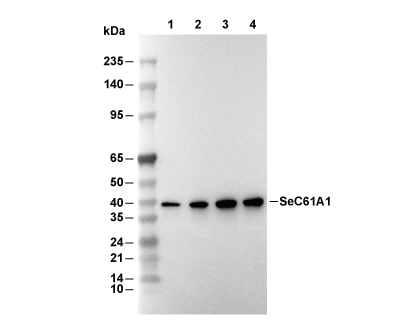 Lane 1: Hela, Lane 2: U-87MG, Lane 3: 3T3, Lane 4: C6