|
Toll Free: (877) 796-6397 -- USA and Canada only -- |
Fax: +1-832-582-8590 Orders: +1-832-582-8158 |
Tech Support: +1-832-582-8158 Ext:3 Please provide your Order Number in the email. |
Technical Data
| Formula | C15H16ClN5 |
||||||
| Molecular Weight | 301.77 | CAS No. | 172889-27-9 | ||||
| Solubility (25°C)* | In vitro | DMSO | 60 mg/mL (198.82 mM) | ||||
| Ethanol | 2 mg/mL (6.62 mM) | ||||||
| Water | Insoluble | ||||||
| In vivo (Add solvents to the product individually and in order) |
|
||||||
|
* <1 mg/ml means slightly soluble or insoluble. * Please note that Selleck tests the solubility of all compounds in-house, and the actual solubility may differ slightly from published values. This is normal and is due to slight batch-to-batch variations. * Room temperature shipping (Stability testing shows this product can be shipped without any cooling measures.) |
|||||||
Preparing Stock Solutions
Biological Activity
| Description | PP2 (AG 1879, AGL 1879), a Src family kinase inhibitor, potently inhibits Lck/Fyn with IC50 of 4 nM/5 nM in cell-free assays, ~100-fold less potent to EGFR, inactive for ZAP-70, JAK2 and PKA. | ||||
|---|---|---|---|---|---|
| Targets |
|
||||
| In vitro | PP2 inhibits Src by binding to an area of the molecule that does not overlap with the ATP binding domain. [2] PP2 (20 μM) induces 40-50% growth inhibition of HT29 cells, this concentration reduces the Src activity as early as 1 hour and maintains a 35% inhibition of Src activity for 2 days. PP2 (100 mM) decreases the Src activity of HT29 cells in a dose-dependent manner. PP2 (1 mM-100 mM) causes a dose-dependent growth inhibition of human colon cancer cells (HT29, SW480, and PMCO1), liver cancer cells (PLC/PRF/5, KYN-2, Li7, and HepG2), and breast cancer cells (MCF-7, MDA-MB-468, and BT-474). PP2 (20 μM) significantly increases aggregation in most of the cancer cells (HT29, SW480, PMCO1, PLC/PRF/5, KYN-2, Li7, MCF-7, and MDA-MB-468) in E-cadherin dependent manner. PP2 (20 μM) enhances E-cadherin expression and also strongly increases E-cadherin’s association with the actin cytoskeleton in cancer cells. PP2 (20 μM) increases the expression of α-catenin, β-catenin, and γ-catenin in HT29 cells, whereas in PLC/PRF/5 and MCF-7 cells, the total protein level of α-catenin does not change, but the levels of β- catenin and γ-catenin increases slightly. [3] PP2 inhibits proliferation of two cervical cancer cells (HeLa and SiHa) in a time- and dose-dependent manner. PP2 (10 μM) down-regulates pSrc-Y416, pEGFR-Y845, and -Y1173 expression levels in HeLa and SiHa cells. PP2 (10 μM) could modulate cell cycle arrest by up-regulating p21(Cip1) and p27(Kip1) in both HeLa and SiHa cells and down-regulating expression of cyclin A, and cyclin dependent kinase-2, -4 (Cdk-2, -4) in HeLa and of cyclin B and Cdk-2 in SiHa. [4] | ||||
| In vivo | PP2 (5 mg/kg/day) induces some slowing in the growth rate of the primary tumors relative to the control treated with vehicle in SCID mice inoculated HT29 cells in the spleen. PP2 (5 mg/kg/day) induces some slowing in the growth rate of the primary tumors relative to the control treated with vehicle in SCID mice inoculated HT29 cells in the spleen. PP2 (5 mg/kg/day) significantly reduces the relative liver weight and liver metastasis volume compared with the controls in SCID mice inoculated HT29 cells in the spleen. [3] PP2 (1.5 mg/kg i.p.) treated rats show approximately 50% reduction of infarct size on T2-weighted MRI and in TTC staining compared with controls in rats with focal ischemic brain injury. PP2 (1.5 mg/kg i.p.) results in better the neurological score than controls in rats with focal ischemic brain injury. [5] |
Protocol (from reference)
| Kinase Assay:[1] |
|
|---|---|
| Cell Assay:[3] |
|
| Animal Study:[3] |
|
References
Customer Product Validation

-
Data from [Data independently produced by , , J Biol Chem, 2014, 289(19): 13026-41 ]

-
Data from [Data independently produced by , , CNS Neurosci Ther, 2017, 23(4):291-300]
-
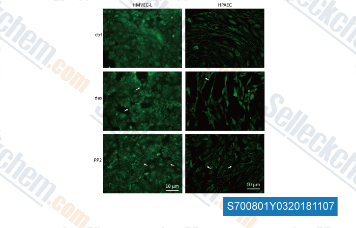
Data from [Data independently produced by , , Front Physiol, 2018, 9: 537]
Selleck's PP2 has been cited by 116 publications
| Microglial P2Y12 Signaling Contributes to Cisplatin-induced Pain Hypersensitivity via IL-18-mediated Central Sensitization in the Spinal Cord [ J Pain, 2023, S1526-5900(23)00013-5] | PubMed: 36646400 |
| LCK facilitates DNA damage repair by stabilizing RAD51 and BRCA1 in the nucleus of chemoresistant ovarian cancer [ J Ovarian Res, 2023, 16(1):122] | PubMed: 37370140 |
| Involvement of NADPH oxidases in the Na/K‑ATPase/Src/ROS oxidant amplification loop in renal fibrosis [ Mol Med Rep, 2023, 28(3)161] | PubMed: 37417374 |
| Phosphatidylserine released from apoptotic cells in tumor induces M2-like macrophage polarization through the PSR-STAT3-JMJD3 axis [ Cancer Commun (Lond), 2022, 42(3):205-222] | PubMed: 35191227 |
| Tankyrase represses autoinflammation through the attenuation of TLR2 signaling [ J Clin Invest, 2022, 132(7)e140869] | PubMed: 35362478 |
| PP2 protects from keratin mutation-associated liver injury and filament disruption via SRC kinase inhibition in male but not female mice [ Hepatology, 2022, 10.1002/hep.32574] | PubMed: 35586977 |
| The LCK-14-3-3ζ-TRPM8 axis regulates TRPM8 function/assembly and promotes pancreatic cancer malignancy [ Cell Death Dis, 2022, 13(6):524] | PubMed: 35665750 |
| Listeria toxin promotes phosphorylation of the inflammasome adaptor ASC through Lyn and Syk to exacerbate pathogen expansion [ Cell Rep, 2022, 38(8):110414] | PubMed: 35196496 |
| The Lectin LecB Induces Patches with Basolateral Characteristics at the Apical Membrane to Promote Pseudomonas aeruginosa Host Cell Invasion [ mBio, 2022, 13(3):e0081922] | PubMed: 35491830 |
| Opposite Effects of Src Family Kinases on YAP and ERK Activation in Pancreatic Cancer Cells: Implications for Targeted Therapy [ Mol Cancer Ther, 2022, 21(11):1652-1662] | PubMed: 35999654 |
RETURN POLICY
Selleck Chemical’s Unconditional Return Policy ensures a smooth online shopping experience for our customers. If you are in any way unsatisfied with your purchase, you may return any item(s) within 7 days of receiving it. In the event of product quality issues, either protocol related or product related problems, you may return any item(s) within 365 days from the original purchase date. Please follow the instructions below when returning products.
SHIPPING AND STORAGE
Selleck products are transported at room temperature. If you receive the product at room temperature, please rest assured, the Selleck Quality Inspection Department has conducted experiments to verify that the normal temperature placement of one month will not affect the biological activity of powder products. After collecting, please store the product according to the requirements described in the datasheet. Most Selleck products are stable under the recommended conditions.
NOT FOR HUMAN, VETERINARY DIAGNOSTIC OR THERAPEUTIC USE.