|
Toll Free: (877) 796-6397 -- USA and Canada only -- |
Fax: +1-832-582-8590 Orders: +1-832-582-8158 |
Tech Support: +1-832-582-8158 Ext:3 Please provide your Order Number in the email. |
Technical Data
| Formula | C16H18N2OS.HBr |
|||
| Molecular Weight | 367.3 | CAS No. | 63208-82-2 | |
| Solubility (25°C)* | In vitro | DMSO | 73 mg/mL (198.74 mM) | |
| Water | Insoluble | |||
| Ethanol | Insoluble | |||
|
* <1 mg/ml means slightly soluble or insoluble. * Please note that Selleck tests the solubility of all compounds in-house, and the actual solubility may differ slightly from published values. This is normal and is due to slight batch-to-batch variations. * Room temperature shipping (Stability testing shows this product can be shipped without any cooling measures.) |
||||
Preparing Stock Solutions
Biological Activity
| Description | Pifithrin-α is an inhibitor of p53, inhibiting p53-dependent transactivation of p53-responsive genes. Pifithrin-α is also a potent agonist of the aryl hydrocarbon receptor (AhR). | |
|---|---|---|
| Targets |
|
|
| In vitro | Pifithrin-α inhibits p53-dependent transactivation of p53-responsive genes in ConA cells. Pifithrin-α (10 μM) inhibits apoptotic death of C8 cells induced by Dox, etoposide, Taxol, cytosine arabinoside. Pifithrin-α inhibits p53-dependent growth arrest of human diploid fibroblasts in response to DNA damage but has no effect on p53-deficient fibroblasts. Pifithrin-α may modulate the nuclear import or export (or both) of p53 or may decrease the stability of nuclear p53. [1] Pifithrin-α (100-200 nM) completely suppresses the camptothecin-induced increase in the level of p53 DNA binding as well as the p53-responsive gene Bax in hippocampal cell. Pifithrin-α also decreases the basal level of p53 DNA-binding activity. Pifithrin-α (200 nM) protects cultured hippocampal neurons against death induced by DNA-damaging agents. Pifithrin-α (200 μM) stabilizes mitochondrial function, suppresses caspase activation and protects cultured hippocampal neurons against death induced by glutamate and amyloid β-peptide. [2] Pifithrin α, in addition to p53, can suppress heat shock and glucocorticoid receptor signaling but has no effect on nuclear factor-kappaB signaling. Pifithrin α (10 μM) reduces activation of heat shock transcription factor (HSF1) and increases cell sensitivity to heat. Pifithrin α (10 μM) reduces activation of glucocorticoid receptor and rescues mouse thymocytes from apoptotic death after dexamethasone treatment in HeLa cells. [3] PFTalpha blocks p53-mediated induction of p21/Waf-1 in human embryonic kidney cells. PFTalpha does, however, cause a left shift in the dexamethasone dose response curve by increasing intracellular dexamethasone concentration. [4] | |
| In vivo | Pifithrin-α (2.2 mg/kg i.p.) treatment completely rescues mice (C57BL and Balb/c) of both strains from 60% killing doses of gamma irradiation (8 Gy for C57BL and 6 Gy for Balb/c). Pifithrin-α-injected mice lost less weight than irradiated mice that are not pretreated with the Pifithrin-α. Pifithrin-α (2.2 mg/kg) abrogates p53-dependent regulation of DNA replication after whole-body gamma irradiation in mice. [1] Pifithrin-α (2 mg/kg i.p.) 30 min prior to middle cerebral artery occlusion treatment of mice reduces ischemic brain injury and protects hippocampal neurons against excitotoxic injury. [2] Pifithrin α (3.6 μg/kg i.p.) inhibits Dex-induced degeneration of the thymus in mice. [3] Pifithrin α (2 mg/kg) results in a significantly lower degree of motor disability in rats receiving transient occlusion of the middle cerebral artery as compared with controls. Pifithrin α-treated animals has less motor disability and smaller infarcts when the drug is administered up to an hour after stroke onset. Pifithrin α results in significantly lower motor disability scores in rats than in the vehicle-treated animals at 7 days post-op. Pifithrin α results in significant reduction of apoptosis in rats as indicated by Tunel and caspase 3 staining. [5] |
Protocol (from reference)
| Cell Assay:[3] |
|
|---|---|
| Animal Study:[1] |
|
References
Customer Product Validation

-
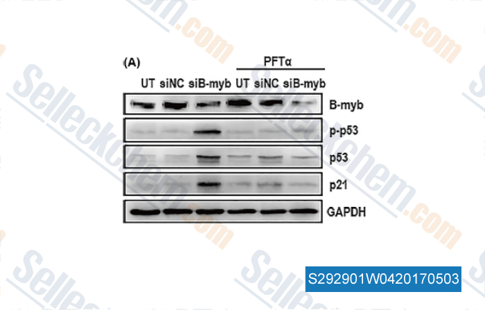
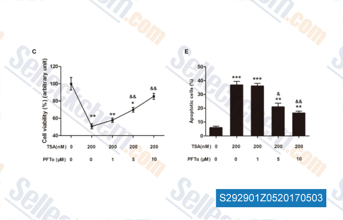
, , Cell Prolif, 2017, doi: 10.1111/cpr.12319

-
Data from [Data independently produced by , , Cell Prolif, 2017, 50(6)]

-
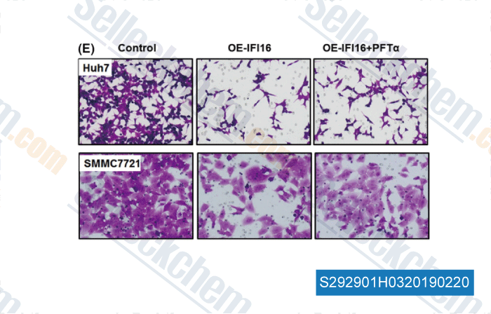
Data from [Data independently produced by , , J Biol Chem, 2016, 291(9):4462-72]

-
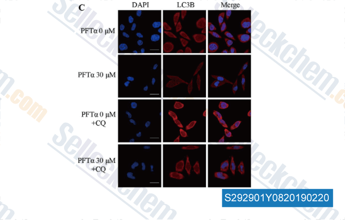
Data from [Data independently produced by , , Int J Biol Sci, 2016, 12(11):1298-1308]
Selleck's Pifithrin-α (PFTα) HBr has been cited by 123 publications
| BOP1 contributes to the activation of autophagy in polycystic ovary syndrome via nucleolar stress response [ Cell Mol Life Sci, 2024, 81(1):101] | PubMed: 38409361 |
| Kidney tubular epithelial cells control interstitial fibroblast fate by releasing TNFAIP8-encapsulated exosomes [ Cell Death Dis, 2023, 14(10):672] | PubMed: 37828075 |
| Suppression of TREX1 deficiency-induced cellular senescence and interferonopathies by inhibition of DNA damage response [ iScience, 2023, 26(7):107090] | PubMed: 37416470 |
| Proximal Tubular Lats2 Ablation Exacerbates Ischemia/Reperfusion Injury -IRI)-Induced Renal Maladaptive Repair through the Upregulation of P53 [ Int J Mol Sci, 2023, 24(20)15258] | PubMed: 37894939 |
| Proximal Tubular Lats2 Ablation Exacerbates Ischemia/Reperfusion Injury (IRI)-Induced Renal Maladaptive Repair through the Upregulation of P53 [ Int J Mol Sci, 2023, 24(20)15258] | PubMed: 37894939 |
| Hepatitis B doubly spliced protein (HBDSP) promotes hepatocellular carcinoma cell apoptosis via ETS1/GATA2/YY1-mediated p53 transcription [ J Virol, 2023, 10.1128/jvi.01087-23] | PubMed: 37929990 |
| DRP1 Inhibition Enhances Venetoclax-Induced Mitochondrial Apoptosis in TP53-Mutated Acute Myeloid Leukemia Cells through BAX/BAK Activation [ Cancers (Basel), 2023, 15(3)745] | PubMed: 36765703 |
| RPS14 promotes the development and progression of glioma via p53 signaling pathway [ Exp Cell Res, 2023, 423(1):113451] | PubMed: 36535509 |
| Curcumin Analog, HO-3867, Induces Both Apoptosis and Ferroptosis via Multiple Mechanisms in NSCLC Cells with Wild-Type p53 [ Evid Based Complement Alternat Med, 2023, 2023:8378581] | PubMed: 36814470 |
| p53 positively regulates the proliferation of hepatic progenitor cells promoted by laminin-521 [ Signal Transduct Target Ther, 2022, 7(1):290] | PubMed: 36042225 |
RETURN POLICY
Selleck Chemical’s Unconditional Return Policy ensures a smooth online shopping experience for our customers. If you are in any way unsatisfied with your purchase, you may return any item(s) within 7 days of receiving it. In the event of product quality issues, either protocol related or product related problems, you may return any item(s) within 365 days from the original purchase date. Please follow the instructions below when returning products.
SHIPPING AND STORAGE
Selleck products are transported at room temperature. If you receive the product at room temperature, please rest assured, the Selleck Quality Inspection Department has conducted experiments to verify that the normal temperature placement of one month will not affect the biological activity of powder products. After collecting, please store the product according to the requirements described in the datasheet. Most Selleck products are stable under the recommended conditions.
NOT FOR HUMAN, VETERINARY DIAGNOSTIC OR THERAPEUTIC USE.
