|
Toll Free: (877) 796-6397 -- USA and Canada only -- |
Fax: +1-832-582-8590 Orders: +1-832-582-8158 |
Tech Support: +1-832-582-8158 Ext:3 Please provide your Order Number in the email. |
Technical Data
| Formula | C34H34F2N4O6 |
|||
| Molecular Weight | 632.65 | CAS No. | 849217-64-7 | |
| Solubility (25°C)* | In vitro | DMSO | 100 mg/mL (158.06 mM) | |
| Water | Insoluble | |||
| Ethanol | Insoluble | |||
|
* <1 mg/ml means slightly soluble or insoluble. * Please note that Selleck tests the solubility of all compounds in-house, and the actual solubility may differ slightly from published values. This is normal and is due to slight batch-to-batch variations. * Room temperature shipping (Stability testing shows this product can be shipped without any cooling measures.) |
||||
Preparing Stock Solutions
Biological Activity
| Description | Foretinib is an ATP-competitive inhibitor of HGFR and VEGFR, mostly for Met (c-Met) and KDR with IC50 of 0.4 nM and 0.9 nM in cell-free assays. Less potent against Ron, Flt-1/3/4, Kit (c-Kit), PDGFRα/β and Tie-2, and little activity to FGFR1 and EGFR. Phase 2. | |||||||||||
|---|---|---|---|---|---|---|---|---|---|---|---|---|
| Targets |
|
|||||||||||
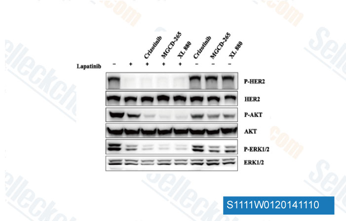
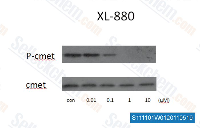
| In vitro | XL880 inhibits HGF receptor family tyrosine kinases with IC50 values of 0.4 nM for Met and 3 nM for Ron. XL880 also inhibits KDR, Flt-1, and Flt-4 with IC50 values of 0.9 nM, 6.8 nM and 2.8 nM, respectively. XL880 inhibits colony growth of B16F10, A549 and HT29 cells with IC50 of 40 nM, 29 nM and 165 nM, respectively. [1] A recent study indicates XL880 affects cell growth differently in gastric cancer cell lines MKN-45 and KATO-III. XL880 inhibits phosphorylation of MET and downstream signaling molecules in MKN-45 cells, while targets GFGR2 in KATO-III cells. [2] | |||||||||||
| In vivo | A single 100 mg/kg oral gavage dose of XL880 results in substantial inhibition of phosphorylation of B16F10 tumor Met and ligand (e.g., HGFor VEGF)-induced receptor phosphorylation of Met in liver and Flk-1/KDR in lung, which both persisted through 24 hours. Treatment with XL880 (30-100 mg/kg, once daily, oral gavage) results in reduction in tumor burden. The lung surface tumor burden is reduced by 50% and 58% following treatment with 30 and 100 mg/kg XL880, respectively. XL880 treatment of mice bearing B16F10 solid tumors also results in dose-dependent tumor growth inhibition of 64% and 87% at 30 and 100 mg/kg, respectively. For both studies, administration of XL880 is well tolerated with no significant body weight loss. [1] XL880 is developed to target abnormal signaling of HGF through Met and simultaneously target several receptors tyrosine kinase involved in tumor angiogenesis. XL880 caused tumor hemorrhage and necrosis in human xenografts within 2 to 4 hours, and maximal tumornecrosis is observed at 96 hours (after five daily doses), resulting in complete regression. [3] |
Protocol (from reference)
| Kinase Assay: |
|
|---|---|
| Cell Assay: |
|
| Animal Study: |
|
References
|
Customer Product Validation
-
Data from [Data independently produced by Cancer Lett, 2013, 340(1), 43-50]
-
, , Dr. Zhang of Tianjin Medical University
-
Data from [Data independently produced by , , Theranostics, 2016, 6(8):1232-43]
-
Data from [Data independently produced by , , Oncotarget, 2018, 9(32):22769-22784]
Selleck's Foretinib has been cited by 94 publications
| Foretinib Is Effective against Triple-Negative Breast Cancer Cells MDA-MB-231 In Vitro and In Vivo by Down-Regulating p-MET/HGF Signaling [ Int J Mol Sci, 2023, 24(1)757] | PubMed: 36614199 |
| A functional sgRNA-CRISPR screening method for generating murine RET and NTRK1 rearranged oncogenes [ Biol Open, 2023, 12(8)bio059994] | PubMed: 37470475 |
| Combined Mcl-1 and YAP1/TAZ inhibition for treatment of metastatic uveal melanoma [ Melanoma Res, 2023, 10.1097/CMR.0000000000000911] | PubMed: 37467061 |
| Adaptive c-Met-PLXDC2 Signaling Axis Mediates Cancer Stem Cell Plasticity to Confer Radioresistance-associated Aggressiveness in Head and Neck Cancer [ Cancer Res Commun, 2023, 3(4):659-671] | PubMed: 37089864 |
| A Rapid, Functional sgRNA Screening Method for Generating Murine RET and NTRK1 Fusion Oncogenes [ bioRxiv, 2023, 2023.04.06.535912] | PubMed: 37066347 |
| A Ctnnb1 enhancer regulates neocortical neurogenesis by controlling the abundance of intermediate progenitors [ Cell Discov, 2022, 8(1):74] | PubMed: 35915089 |
| Phase separation of insulin receptor substrate 1 drives the formation of insulin/IGF-1 signalosomes [ Cell Discov, 2022, 8(1):60] | PubMed: 35764611 |
| TMUB1 is an endoplasmic reticulum-resident escortase that promotes the p97-mediated extraction of membrane proteins for degradation [ Mol Cell, 2022, S1097-2765(22)00663-3] | PubMed: 35961308 |
| A community challenge for a pancancer drug mechanism of action inference from perturbational profile data [ Cell Rep Med, 2022, 3(1):100492] | PubMed: 35106508 |
| PDGF signaling inhibits mitophagy in glioblastoma stem cells through N6-methyladenosine [ Dev Cell, 2022, 57(12):1466-1481.e6] | PubMed: 35659339 |
RETURN POLICY
Selleck Chemical’s Unconditional Return Policy ensures a smooth online shopping experience for our customers. If you are in any way unsatisfied with your purchase, you may return any item(s) within 7 days of receiving it. In the event of product quality issues, either protocol related or product related problems, you may return any item(s) within 365 days from the original purchase date. Please follow the instructions below when returning products.
SHIPPING AND STORAGE
Selleck products are transported at room temperature. If you receive the product at room temperature, please rest assured, the Selleck Quality Inspection Department has conducted experiments to verify that the normal temperature placement of one month will not affect the biological activity of powder products. After collecting, please store the product according to the requirements described in the datasheet. Most Selleck products are stable under the recommended conditions.
NOT FOR HUMAN, VETERINARY DIAGNOSTIC OR THERAPEUTIC USE.