-
 Australia
-
 Austria
-
 Belgium
-
 Brazil
-
 Canada
-
 China
China
-
 Czech Republic
-
 Denmark
-
 Finland
-
 France
-
 Germany
-
 Greece
-
 Hong Kong
-
 Hungary
-
 Iceland
-
 India
India
-
 Ireland
-
 Israel
-
 Italy
Italy
-
 Japan
Japan
-
 Korea
Korea
-
 Luxembourg
-
 Malaysia
-
 Netherlands
-
 New Zealand
-
 Norway
-
 Poland
-
 Qatar
Qatar
-
 Romania
-
 Saudi Arabia
-
 Singapore
-
 Spain
Spain
-
 Sweden
-
 Switzerland
-
 Taiwan
-
 Turkey
-
 United Kingdom
-
 United States
research use only
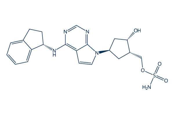
MLN4924 (Pevonedistat) NAE inhibitor
Cat.No.S7109
Quality Control
| Related Targets | Proteasome E3 Ligase DUB SUMO p97 E2 conjugating |
|---|---|
| Other E1 Activating Inhibitors | TAK-243 (MLN7243) PYR-41 ML792 Pevonedistat hydrochloride TAS4464 COH000 DKM 2-93 PYZD-4409 NEDD8 inhibitor M22 |
Solubility in DMSO or Water
|
In vitro |
DMSO
: 89 mg/mL
(200.66 mM)
Ethanol : 22 mg/mL Water : Insoluble The solubility data above are all experimental results (not literature values). |
Molarity Calculator
|
In vivo |
|||||
In vivo Formulation Calculator (Clear solution)
Step 1: Enter information below (Recommended: An additional animal making an allowance for loss during the experiment)
Step 2: Enter the in vivo formulation (This is only the calculator, not formulation. Please contact us first if there is no in vivo formulation at the solubility Section.)
Calculation results:
Working concentration: mg/ml;
Method for preparing DMSO master liquid: mg drug pre-dissolved in μL DMSO ( Master liquid concentration mg/mL, Please contact us first if the concentration exceeds the DMSO solubility of the batch of drug. )
Method for preparing in vivo formulation: Take μL DMSO master liquid, next addμL PEG300, mix and clarify, next addμL Tween 80, mix and clarify, next add μL ddH2O, mix and clarify.
Method for preparing in vivo formulation: Take μL DMSO master liquid, next add μL Corn oil, mix and clarify.
Note: 1. Please make sure the liquid is clear before adding the next solvent.
2. Be sure to add the solvent(s) in order. You must ensure that the solution obtained, in the previous addition, is a clear solution before proceeding to add the next solvent. Physical methods such
as vortex, ultrasound or hot water bath can be used to aid dissolving.
Read more about MLN4924 (Pevonedistat) solubility in DMSO or water
Working Concentrations for Cell Culture Treatment
Step 1: Select a research area to find working concentrations, or search directly for a cell line
Step 2: Select a cell line to find matched working concentrations
Read more about MLN4924 (Pevonedistat) working concentrations for cell culture treatment
Working Concentrations for Animal Model Treatment
Step 1: Select a research area to find working concentrations, or search directly for an animal type
Step 2: Select an animal type to find matched working concentrations
Mechanism of Action
| Information | MLN4924 (Pevonedistat) is a potent NEDD8-activating enzyme (NAE) inhibitor. It affects PI3K/AKT, NF-κB, and RhoA/ROCK signaling pathways by blocking Cullin-RING ligase neddylation and stabilizing substrates like p21 and p27, thereby inhibiting cancer cell proliferation, migration, and inducing apoptosis. |
|---|---|
|
Read more about MLN4924 (Pevonedistat) IC50 for cell-based and cell-free assays |
|
| Primary Applications and Mechanism of Action | |
Chemical Information, Storage & Stability
| Molecular Weight | 443.52 | Formula | C21H25N5O4S |
Storage (From the date of receipt) | |
|---|---|---|---|---|---|
| CAS No. | 905579-51-3 | Download SDF | Storage of Stock Solutions |
|
|
Tech Support
Tel: +1-832-582-8158 Ext:3
If you have any other enquiries, please leave a message.
Products are for research use only. Not for human use. We do not sell to patients.
©Copyright 2013 Selleck Chemicals. All Rights Reserved.