|
How to Cite 1. For In-Text Citation (Materials & Methods): 2. For Key Resources Table: |
||
|
Toll Free: (877) 796-6397 -- USA and Canada only -- |
Fax: +1-832-582-8590 Orders: +1-832-582-8158 |
Tech Support: +1-832-582-8158 Ext:3 Please provide your Order Number in the email. We strive to reply to |
Biological Description
| Specificity | VCP Antibody [P13E12] detects endogenous levels of total VCP protein. |
|---|---|
| Background | VCP (p97), a type II AAA+ ATPase abundantly expressed across tissues, assembles into homo-hexameric double-ring cylinders that extract ubiquitinated client proteins from membranes, chromatin, ribosomes, and protein complexes for proteasomal degradation, autophagy, or refolding. Each protomer features an N-domain (ubiquitin-binding UBX-like fold coordinating cofactors via S2/S3/S4 subdomains), D1/D2 ATPase domains (RecA-like folds with Walker A/B motifs, with D2 providing dominant hydrolysis and generating ~100 nm translocation force), L1/L2 interdomain linkers (for allosteric regulation), and a C-terminal tail (conferring cofactor specificity); the hexamer’s central pore, lined with 48 aromatic/hydrophobic residues, threads unfolded polypeptides. It exhibits segregase activity: ATP binding to the D1 ring assembles the hexamer, while D2 hydrolysis generates power strokes that sequentially grip clients via N-domain–cofactor complexes (UFD1-NPL4/VCIP135/p37), pulling substrates through the central pore (~1–2 nm/sec unfolding rate) for ER-associated degradation (ERAD of ubiquitinated misfolded proteins), ribosome quality control (removal of stalled nascent chains), endosomal-lysosomal damage repair (ELDR/lysophagy), and mitotic spindle disassembly (Aurora A chromatin eviction); phosphorylation (Ser784 by ATM, Thr688 by CDK) modulates VCP localization. VCP enforces proteostasis (stress granule clearance), organelle dynamics (mitochondrial fusion/fission), DNA repair (HRR/NHEJ), and cell cycle fidelity (G2/M checkpoint); its enrichment in nervous tissue explains multisystem proteinopathies. Disease relevance: heterozygous mutations (R93C/R155C) cause IBMPFD2, ALS14, and multisystem proteinopathy via toxic gain-of-function (constitutive hexamerization, ~2-fold hyperATPase activity). |
Usage Information
| Application | WB | Dilution |
|
||
|---|---|---|---|---|---|
| Reactivity | Human, Mouse, Rat, Monkey | ||||
| Source | Rabbit Monoclonal Antibody | MW | 89 kDa | ||
| Storage Buffer | PBS, pH 7.2+50% Glycerol+0.05% BSA+0.01% NaN3 | Storage (from the date of receipt) |
-20°C (avoid freeze-thaw cycles), 2 years | ||
| WB |
Experimental Protocol:
Sample preparation
1. Tissue: Lyse the tissue sample by adding an appropriate volume of ice-cold RIPA/NP-40 Lysis Buffer (containing Protease Inhibitor Cocktail),and homogenize the tissue at a low temperature. 2. Adherent cell: Aspirate the culture medium and wash the cells with ice-cold PBS twice. Lyse the cells by adding an appropriate volume of RIPA/NP-40 Lysis Buffer (containing Protease Inhibitor Cocktail) and put the sample on ice for 5 min. 3. Suspension cell: Transfer the culture medium to a pre-cooled centrifuge tube. Centrifuge and aspirate the supernatant. Wash the cells with ice-cold PBS twice. Lyse the cells by adding an appropriate volume of RIPA/NP-40 Lysis Buffer (containing Protease Inhibitor Cocktail) and put the sample on ice for 5 min. 4. Place the lysate into a pre-cooled microcentrifuge tube. Centrifuge at 4°C for 15 min. Collect the supernatant;
5. Remove a small volume of lysate to determine the protein concentration;
6. Combine the lysate with protein loading buffer. Boil 20 µL sample under 95-100°C for 5 min. Centrifuge for 5 min after cool down on ice.
2. Power up 80V for 30 minutes. Then the power supply is adjusted (110 V~150 V), the Marker is observed, and the electrophoresis can be stopped when the indicator band of the predyed protein Marker where the protein is located is properly separated. (Note that the current should not be too large when electrophoresis, too large current (more than 150 mA) will cause the temperature to rise, affecting the result of running glue. If high currents cannot be avoided, an ice bath can be used to cool the bath.)
Transfer membrane
1. Take out the converter, soak the clip and consumables in the pre-cooled converter;
2. Activate PVDF membrane with methanol for 1 min and rinse with transfer buffer;
3. Install it in the order of "black edge of clip - sponge - filter paper - filter paper - glue -PVDF membrane - filter paper - filter paper - sponge - white edge of clip"; 4. The protein was electrotransferred to PVDF membrane. ( 0.45 µm PVDF membrane is recommended ) Reference Table for Selecting PVDF Membrane Pore Size Specifications Recommended conditions for wet transfer: 200 mA, 120 min. ( Note that the transfer conditions can be adjusted according to the protein size. For high-molecular-weight proteins, a higher current and longer transfer time are recommended. However, ensure that the transfer tank remains at a low temperature to prevent gel melting.)
Block
1. After electrotransfer, wash the film with TBST at room temperature for 5 minutes;
2. Incubate the film in the blocking solution for 1 hour at room temperature;
3. Wash the film with TBST for 3 times, 5 minutes each time.
Antibody incubation
1. Use 5% skim milk powder to prepare the primary antibody working liquid (recommended dilution ratio for primary antibody 1:1000), gently shake and incubate with the film at 4°C overnight; 2. Wash the film with TBST 3 times, 5 minutes each time;
3. Add the secondary antibody to the blocking solution and incubate with the film gently at room temperature for 1 hour;
4. After incubation, wash the film with TBST 3 times for 5 minutes each time.
Antibody staining
1. Add the prepared ECL luminescent substrate (or select other color developing substrate according to the second antibody) and mix evenly;
2. Incubate with the film for 1 minute, remove excess substrate (keep the film moist), wrap with plastic film, and expose in the imaging system.
|
References
|
Application Data
WB
Validated by Selleck
-
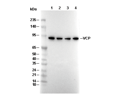 Lane 1: Hela, Lane 2: NIH/3T3, Lane 3: C6, Lane 4: COS