
|
How to Cite 1. For In-Text Citation (Materials & Methods): 2. For Key Resources Table: |
||
|
Toll Free: (877) 796-6397 -- USA and Canada only -- |
Fax: +1-832-582-8590 Orders: +1-832-582-8158 |
Tech Support: +1-832-582-8158 Ext:3 Please provide your Order Number in the email. We strive to reply to |
Biological Description
| Specificity | Toll-like Receptor 4 Antibody (Rabbit mAb) [D2G12] detects endogenous levels of total Toll-like Receptor 4 protein. |
|---|---|
| Background | Toll-like receptor 4 (TLR4), also known as CD284, is a pattern-recognition receptor of the Toll-like receptor family that senses pathogen- and damage-associated molecular patterns at the plasma membrane and in endosomal compartments, providing a primary trigger for innate immune activation against Gram‑negative bacteria and sterile injury. The receptor has a large extracellular leucine-rich repeat domain that binds lipopolysaccharide (LPS) in complex with the co-receptor MD‑2 and accessory protein CD14, a single transmembrane helix, and a cytoplasmic TIR domain that engages downstream adaptor proteins to assemble signaling signalosomes. LPS-binding protein transfers monomeric LPS to CD14, which delivers LPS to the TLR4–MD‑2 ectodomain; binding induces homodimerization of the LPS–MD‑2–TLR4 complex and brings the intracellular TIR domains into proximity, creating a scaffold for recruitment of adaptor molecules. At the plasma membrane, TIRAP (Mal) and MyD88 are recruited to the activated TIR domains to form a MyD88-templated “myddosome” that engages IRAK kinases and TRAF6, leading to activation of the IKK complex, phosphorylation and degradation of IκB, and nuclear translocation of NF‑κB, together with activation of MAPKs including p38, ERK and JNK. This MyD88-dependent arm drives early transcription of proinflammatory cytokines, chemokines, and costimulatory molecules, establishing a rapid inflammatory and immune-priming response. After initial signaling, TLR4 is internalized into early endosomes where TRAM and TRIF act as sorting adaptors, assembling a distinct TRIF-dependent complex that activates TBK1 and IKKε, phosphorylates IRF3 and induces type I interferon and interferon-stimulated gene expression, providing an antiviral and immunoregulatory dimension to TLR4 signaling that is spatially segregated from the plasma membrane proinflammatory output. Clustered TLR4–MyD88 assemblies template helical myddosome formation and position TRAF3 and TRAF6, whose partially overlapping binding sites on MyD88 integrate anti-viral/anti-inflammatory (TRAF3) versus proinflammatory (TRAF6) outputs, highlighting how TLR4 pathway branching is encoded at the adaptor and signalosome level. TLR4 recognizes not only LPS but also endogenous danger ligands such as HMGB1, heat shock proteins, and oxidized lipoproteins, and ligation by these DAMPs contributes to noninfectious inflammation in conditions including atherosclerosis, diabetes, neurodegeneration, and tissue injury. In sepsis, extremely low concentrations of endotoxin–MD‑2 complexes are sufficient to drive strong TLR4‑dependent cell activation, and uncontrolled MyD88 and TRIF signaling amplify cytokine storms and coagulation pathways, making TLR4 a central node in septic pathophysiology and a target for antagonists that disrupt LPS–MD‑2–TLR4 interactions or downstream adaptor recruitment. In atherosclerosis and vascular disease, TLR4 expressed on endothelial cells, smooth muscle cells and macrophages responds to modified lipoproteins and other endogenous ligands, promoting NF‑κB‑driven expression of adhesion molecules, chemokines and matrix-degrading enzymes that support plaque formation and instability. |
Usage Information
| Application | WB | Dilution |
|
||
|---|---|---|---|---|---|
| Reactivity | Mouse | ||||
| Source | Rabbit Monoclonal Antibody | MW | 96 kDa | ||
| Storage Buffer | PBS, pH 7.2+50% Glycerol+0.05% BSA+0.01% NaN3 | Storage (from the date of receipt) |
-20°C (avoid freeze-thaw cycles), 2 years | ||
| WB |
Experimental Protocol:
Sample preparation
1. Tissue: Lyse the tissue sample by adding an appropriate volume of ice-cold RIPA/NP-40 Lysis Buffer (containing Protease Inhibitor Cocktail),and homogenize the tissue at a low temperature or lyse it by sonication on ice, then incubate on ice for 30 minutes. 2. Adherent cell: Aspirate the culture medium and wash the cells with ice-cold PBS twice. Lyse the cells by adding an appropriate volume of RIPA/NP-40 Lysis Buffer (containing Protease Inhibitor Cocktail) , sonicate to lyse the cells, and incubate on ice for 30 minutes. 3. Suspension cell: Transfer the culture medium to a pre-cooled centrifuge tube. Centrifuge and aspirate the supernatant. Wash the cells with ice-cold PBS twice. Lyse the cells by adding an appropriate volume of RIPA/NP-40 Lysis Buffer (containing Protease Inhibitor Cocktail) , sonicate to lyse the cells, and incubate on ice for 30 minutes. 4. Place the lysate into a pre-cooled microcentrifuge tube. Centrifuge at 4°C for 15 min. Collect the supernatant;
5. Remove a small volume of lysate to determine the protein concentration;
6. Combine the lysate with protein loading buffer. Boil 20 µL sample under 95-100°C for 5 min. Centrifuge for 5 min after cool down on ice.
Electrophoretic separation
1. According to the concentration of extracted protein, load appropriate amount of protein sample and marker onto SDS-PAGE gels for electrophoresis. Recommended separating gel (lower gel) concentration: 10%. Reference Table for Selecting SDS-PAGE Separation Gel Concentrations 2. Power up 80V for 30 minutes. Then the power supply is adjusted (110 V~150 V), the Marker is observed, and the electrophoresis can be stopped when the indicator band of the predyed protein Marker where the protein is located is properly separated. (Note that the current should not be too large when electrophoresis, too large current (more than 150 mA) will cause the temperature to rise, affecting the result of running glue. If high currents cannot be avoided, an ice bath can be used to cool the bath.)
Transfer membrane
1. Take out the converter, soak the clip and consumables in the pre-cooled converter;
2. Activate PVDF membrane with methanol for 1 min and rinse with transfer buffer;
3. Install it in the order of "black edge of clip - sponge - filter paper - filter paper - glue -PVDF membrane - filter paper - filter paper - sponge - white edge of clip"; 4. The protein was electrotransferred to PVDF membrane. ( 0.45 µm PVDF membrane is recommended ) Reference Table for Selecting PVDF Membrane Pore Size Specifications Recommended conditions for wet transfer: 200 mA, 120 min. ( Note that the transfer conditions can be adjusted according to the protein size. For high-molecular-weight proteins, a higher current and longer transfer time are recommended. However, ensure that the transfer tank remains at a low temperature to prevent gel melting.)
Block
1. After electrotransfer, wash the film with TBST at room temperature for 5 minutes;
2. Incubate the film in the blocking solution for 1 hour at room temperature;
3. Wash the film with TBST for 3 times, 5 minutes each time.
Antibody incubation
1. Use primary antibody dilution buffer to prepare the primary antibody working liquid (recommended dilution ratio for primary antibody 1:1000), gently shake and incubate with the film at 4°C overnight; 2. Wash the film with TBST 3 times, 5 minutes each time;
3. Add the secondary antibody to the blocking solution and incubate with the film gently at room temperature for 1 hour;
4. After incubation, wash the film with TBST 3 times for 5 minutes each time.
Antibody staining
1. Add the prepared ECL luminescent substrate (or select other color developing substrate according to the second antibody) and mix evenly;
2. Incubate with the film for 1 minute, remove excess substrate (keep the film moist), wrap with plastic film, and expose in the imaging system.
|
References
|
Application Data
WB
Validated by Selleck
-
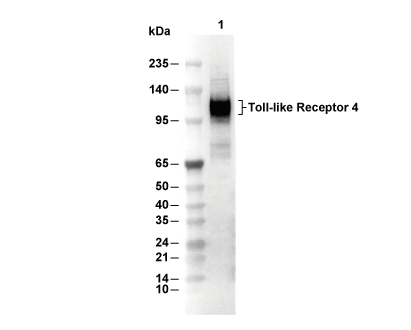 Lane 1: 3T3