|
Toll Free: (877) 796-6397 -- USA and Canada only -- |
Fax: +1-832-582-8590 Orders: +1-832-582-8158 |
Tech Support: +1-832-582-8158 Ext:3 Please provide your Order Number in the email. |
Biological Description
| Specificity | TIMP2 Mouse mAb detects endogenous levels of total TIMP2 protein. |
|---|---|
| Background | Tissue inhibitor of metalloproteinases 2 (TIMP2) is a member of the TIMP family that regulates extracellular matrix (ECM) turnover by inhibiting matrix metalloproteinases (MMPs). The TIMP2 gene is located on chromosome 17q25.3 and encodes a 194-amino acid protein with a molecular mass of approximately 21 kDa. It is widely expressed in most cell types and is the most commonly observed TIMP in normal tissues.TIMP2 plays a crucial role in controlling pericellular proteolysis, impacting processes like cell growth, proliferation, migration, inflammation, inhibition of cellular invasion, tumorigenesis, metastasis, angiogenesis, and cellular aging. TIMP2 forms high-affinity complexes with various MMPs (e.g., MMP1, MMP2, MMP9), particularly binding to MMP14 (MT1-MMP) on the cell surface, which serves as an adaptor to activate pro-MMP2, leading to subsequent MMP2 and MMP9 activation. Additionally, TIMP2 interacts with α3β1 integrin, promoting cell cycle arrest via SHP-1 activation and nuclear localization of p27. |
Usage Information
| Application | IHC | Dilution |
|
||
|---|---|---|---|---|---|
| Reactivity | Human | ||||
| Source | Mouse | MW | 24 kDa | ||
| Storage Buffer | PBS, pH 7.2+50% Glycerol+0.05% BSA+0.01% NaN₃ | Storage (from the date of receipt) |
–20°C (avoid freeze-thaw cycles), 2 years | ||
| IHC |
Experimental Protocol: Deparaffinization/Rehydration
1. Deparaffinize/hydrate sections:
2. Incubate sections in three washes of xylene for 5 min each.
3. Incubate sections in two washes of 100% ethanol for 10 min each.
4. Incubate sections in two washes of 95% ethanol for 10 min each.
5. Wash sections two times in dH2O for 5 min each.
6.Antigen retrieval: For Citrate: Heat slides in a microwave submersed in 1X citrate unmasking solution until boiling is initiated; continue with 10 min at a sub-boiling temperature (95°-98°C). Cool slides on bench top for 30 min.
Staining
1. Wash sections in dH2O three times for 5 min each.
2. Incubate sections in 3% hydrogen peroxide for 10 min.
3. Wash sections in dH2O two times for 5 min each.
4. Wash sections in wash buffer for 5 min.
5. Block each section with 100–400 µl of blocking solution for 1 hr at room temperature.
6. Remove blocking solution and add 100–400 µl primary antibody diluent in to each section. Incubate overnight at 4°C.
7. Remove antibody solution and wash sections with wash buffer three times for 5 min each.
8. Cover section with 1–3 drops HRPas needed. Incubate in a humidified chamber for 30 min at room temperature.
9. Wash sections three times with wash buffer for 5 min each.
10. Add DAB Chromogen Concentrate to DAB Diluent and mix well before use.
11. Apply 100–400 µl DAB to each section and monitor closely. 1–10 min generally provides an acceptable staining intensity.
12. Immerse slides in dH2O.
13. If desired, counterstain sections with hematoxylin.
14. Wash sections in dH2O two times for 5 min each.
15. Dehydrate sections: Incubate sections in 95% ethanol two times for 10 sec each; Repeat in 100% ethanol, incubating sections two times for 10 sec each; Repeat in xylene, incubating sections two times for 10 sec each.
16. Mount sections with coverslips and mounting medium.
|
Application Data
IHC
Validated by Selleck
-
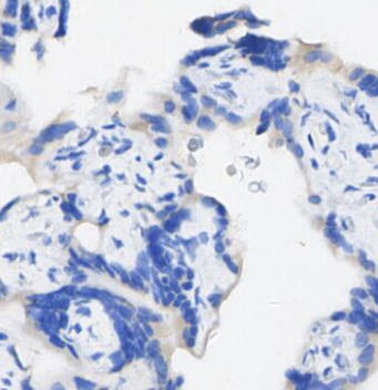 Immunohistochemical analysis of formalin fixed paraffin embedded Human placenta tissue with F1218 at 1/2400 dilution.