|
How to Cite 1. For In-Text Citation (Materials & Methods): 2. For Key Resources Table: |
||
|
Toll Free: (877) 796-6397 -- USA and Canada only -- |
Fax: +1-832-582-8590 Orders: +1-832-582-8158 |
Tech Support: +1-832-582-8158 Ext:3 Please provide your Order Number in the email. We strive to reply to |
Biological Description
| Specificity | TGIF Antibody [C15J1] detects endogenous levels of total TGIF protein. |
|---|---|
| Background | TGIF1 (Homeobox protein TGIF1, 5'-TG-3' interacting factor 1) belongs to the three amino acid loop extension (TALE) homeodomain family of atypical homeobox transcription factors that repress gene expression during embryonic development and cellular differentiation. TGIF1 spans 237 amino acids with a central TALE homeodomain (HD, residues 80-140) featuring a three-residue insertion between helices 1 and 2, enabling recognition of the atypical 5'-TGACA-3' DNA motif through combined major groove contacts by the HD core and minor groove compression by the N-terminal arm. A C-terminal domain (residues 150-237) mediates protein-protein interactions, remaining largely unstructured to flexibly engage partners. TGIF1 docks onto Smad2-Smad4 complexes activated by TGF-β/Nodal signaling, with the HD binding Smad MH1 domains to sterically block DNA access while the C-terminus grips Smad MH2, displacing the complex from promoters and terminating transcription of mesendodermal and ventral midline genes. This active repression bifurcates the forebrain and patterns ventral structures by antagonizing nodal-induced Shh expression in Holoprosencephaly (HPE). TGIF1 also inhibits RXRα transactivation from retinoic acid-responsive elements like CRBPII-RXRE, modulating vitamin A signaling in neural tube closure. In adult cells, TGIF1 curbs epithelial-mesenchymal transition via Smad3 sequestration and cooperates with HDAC1/NCoR to silence pluripotency genes during differentiation. Heterozygous TGIF1 mutations underlie 1-4% of HPE cases, causing holoprosencephaly through haploinsufficiency that disrupts midline signaling and elevates nodal targets. Overexpression drives breast cancer invasion via Twist1 upregulation and glioma proliferation through EGFR pathways. TGIF1 deficiency impairs primitive streak formation and definitive endoderm specification in embryoid bodies. Polyalanine expansions destabilize the protein, linking to richer HPE phenotypes. |
Usage Information
| Application | WB, IP | Dilution |
|
||||
|---|---|---|---|---|---|---|---|
| Reactivity | Human | ||||||
| Source | Rabbit Monoclonal Antibody | MW | 43 kDa | ||||
| Storage Buffer | PBS, pH 7.2+50% Glycerol+0.05% BSA+0.01% NaN3 | Storage (from the date of receipt) |
-20°C (avoid freeze-thaw cycles), 2 years | ||||
| WB |
Experimental Protocol:
Sample preparation
1. Tissue: Lyse the tissue sample by adding an appropriate volume of ice-cold RIPA/Nuclear Lysis Buffer (containing Protease Inhibitor Cocktail),and homogenize the tissue at a low temperature. 2. Adherent cell: Aspirate the culture medium and wash the cells with ice-cold PBS twice. Lyse the cells by adding an appropriate volume of RIPA/Nuclear Lysis Buffer (containing Protease Inhibitor Cocktail) and put the sample on ice for 5 min. 3. Suspension cell: Transfer the culture medium to a pre-cooled centrifuge tube. Centrifuge and aspirate the supernatant. Wash the cells with ice-cold PBS twice. Lyse the cells by adding an appropriate volume of RIPA/Nuclear Lysis Buffer (containing Protease Inhibitor Cocktail) and put the sample on ice for 5 min. 4. Place the lysate into a pre-cooled microcentrifuge tube. Centrifuge at 4°C for 15 min. Collect the supernatant;
5. Remove a small volume of lysate to determine the protein concentration;
6. Combine the lysate with protein loading buffer. Boil 20 µL sample under 95-100°C for 5 min. Centrifuge for 5 min after cool down on ice.
2. Power up 80V for 30 minutes. Then the power supply is adjusted (110 V~150 V), the Marker is observed, and the electrophoresis can be stopped when the indicator band of the predyed protein Marker where the protein is located is properly separated. (Note that the current should not be too large when electrophoresis, too large current (more than 150 mA) will cause the temperature to rise, affecting the result of running glue. If high currents cannot be avoided, an ice bath can be used to cool the bath.)
Transfer membrane
1. Take out the converter, soak the clip and consumables in the pre-cooled converter;
2. Activate PVDF membrane with methanol for 1 min and rinse with transfer buffer;
3. Install it in the order of "black edge of clip - sponge - filter paper - filter paper - glue -PVDF membrane - filter paper - filter paper - sponge - white edge of clip"; 4. The protein was electrotransferred to PVDF membrane. ( 0.45 µm PVDF membrane is recommended ) Reference Table for Selecting PVDF Membrane Pore Size Specifications Recommended conditions for wet transfer: 200 mA, 120 min. ( Note that the transfer conditions can be adjusted according to the protein size. For high-molecular-weight proteins, a higher current and longer transfer time are recommended. However, ensure that the transfer tank remains at a low temperature to prevent gel melting.)
Block
1. After electrotransfer, wash the film with TBST at room temperature for 5 minutes;
2. Incubate the film in the blocking solution for 1 hour at room temperature;
3. Wash the film with TBST for 3 times, 5 minutes each time.
Antibody incubation
1. Use 5% skim milk powder to prepare the primary antibody working liquid (recommended dilution ratio for primary antibody 1:5000), gently shake and incubate with the film at 4°C overnight; 2. Wash the film with TBST 3 times, 5 minutes each time;
3. Add the secondary antibody to the blocking solution and incubate with the film gently at room temperature for 1 hour;
4. After incubation, wash the film with TBST 3 times for 5 minutes each time.
Antibody staining
1. Add the prepared ECL luminescent substrate (or select other color developing substrate according to the second antibody) and mix evenly;
2. Incubate with the film for 1 minute, remove excess substrate (keep the film moist), wrap with plastic film, and expose in the imaging system.
|
References
|
Application Data
WB
Validated by Selleck
-
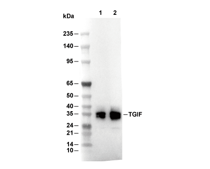 Lane 1: Hela, Lane 2: K562