
|
How to Cite 1. For In-Text Citation (Materials & Methods): 2. For Key Resources Table: |
||
|
Toll Free: (877) 796-6397 -- USA and Canada only -- |
Fax: +1-832-582-8590 Orders: +1-832-582-8158 |
Tech Support: +1-832-582-8158 Ext:3 Please provide your Order Number in the email. We strive to reply to |
Biological Description
| Specificity | TDRD3 Antibody (Rabbit mAb) [C20J18] detects endogenous levels of total TDRD3 protein. |
|---|---|
| Background | TDRD3 is a multidomain Tudor family protein that functions as a reader of asymmetric dimethyl‑arginine marks and links arginine methylation to transcriptional control, R‑loop metabolism, and RNA stress–response compartments. The protein contains an N‑terminal ubiquitin‑associated domain, a central Tudor domain with a defined methyl‑arginine–binding surface, and additional low‑complexity and nucleic acid–recognition regions that support scaffolding of protein and RNA complexes. The Tudor domain recognizes asymmetric dimethyl‑arginine on histone H3 Arg17 and histone H4 Arg3, marks written mainly by PRMT1 and associated with transcriptional activation, and this binding recruits TDRD3 to active chromatin at promoters and enhancers where arginine‑methylated tails are present. At these loci, TDRD3 acts as a coactivator by bringing in additional factors such as Topoisomerase IIIβ and helicases including DHX9, which relax negatively supercoiled DNA and resolve R‑loops that arise during high transcriptional output, thereby supporting continued elongation and limiting exposure of persistent DNA–RNA hybrids. Direct interaction between the TDRD3 Tudor domain and DHX9 guides DHX9 to specific promoters, and loss of TDRD3 reduces DHX9 chromatin occupancy and increases promoter‑proximal R‑loop accumulation, illustrating a defined pathway in which methyl‑histone reading and helicase recruitment converge on genome stability maintenance at transcribed genes. TDRD3 also localizes to translating polyribosomes and accumulates in cytoplasmic stress granules upon diverse stress stimuli; in these structures, it binds dimethyl‑arginine–containing RNA‑binding proteins, including G3BP1, via its Tudor domain and promotes stress granule assembly by enhancing RNA binding and increasing the valence of protein–RNA interaction networks that drive liquid–liquid phase separation. PRMT1 methylates key stress granule RBPs on RGG motifs, and TDRD3 acts as the methyl‑mark reader in this writer–reader pair, so coordinated PRMT1–TDRD3 activity regulates the threshold and dynamics of stress granule formation and influences how translationally stalled mRNAs are partitioned during stress. |
Usage Information
| Application | WB, IP | Dilution |
|
||||
|---|---|---|---|---|---|---|---|
| Reactivity | Human, Mouse, Rat, Monkey | ||||||
| Source | Rabbit Monoclonal Antibody | MW | 83 kDa | ||||
| Storage Buffer | PBS, pH 7.2+50% Glycerol+0.05% BSA+0.01% NaN3 | Storage (from the date of receipt) |
-20°C (avoid freeze-thaw cycles), 2 years | ||||
| WB |
Experimental Protocol:
Sample preparation
1. Tissue: Lyse the tissue sample by adding an appropriate volume of ice-cold RIPA/NP-40 Lysis Buffer (containing Protease Inhibitor Cocktail),and homogenize the tissue at a low temperature or lyse it by sonication on ice, then incubate on ice for 30 minutes. 2. Adherent cell: Aspirate the culture medium and wash the cells with ice-cold PBS twice. Lyse the cells by adding an appropriate volume of RIPA/NP-40 Lysis Buffer (containing Protease Inhibitor Cocktail) , sonicate to lyse the cells, and incubate on ice for 30 minutes. 3. Suspension cell: Transfer the culture medium to a pre-cooled centrifuge tube. Centrifuge and aspirate the supernatant. Wash the cells with ice-cold PBS twice. Lyse the cells by adding an appropriate volume of RIPA/NP-40 Lysis Buffer (containing Protease Inhibitor Cocktail) , sonicate to lyse the cells, and incubate on ice for 30 minutes. 4. Place the lysate into a pre-cooled microcentrifuge tube. Centrifuge at 4°C for 15 min. Collect the supernatant;
5. Remove a small volume of lysate to determine the protein concentration;
6. Combine the lysate with protein loading buffer. Boil 20 µL sample under 95-100°C for 5 min. Centrifuge for 5 min after cool down on ice.
Electrophoretic separation
1. According to the concentration of extracted protein, load appropriate amount of protein sample and marker onto SDS-PAGE gels for electrophoresis. Recommended separating gel (lower gel) concentration: 10%. Reference Table for Selecting SDS-PAGE Separation Gel Concentrations 2. Power up 80V for 30 minutes. Then the power supply is adjusted (110 V~150 V), the Marker is observed, and the electrophoresis can be stopped when the indicator band of the predyed protein Marker where the protein is located is properly separated. (Note that the current should not be too large when electrophoresis, too large current (more than 150 mA) will cause the temperature to rise, affecting the result of running glue. If high currents cannot be avoided, an ice bath can be used to cool the bath.)
Transfer membrane
1. Take out the converter, soak the clip and consumables in the pre-cooled converter;
2. Activate PVDF membrane with methanol for 1 min and rinse with transfer buffer;
3. Install it in the order of "black edge of clip - sponge - filter paper - filter paper - glue -PVDF membrane - filter paper - filter paper - sponge - white edge of clip"; 4. The protein was electrotransferred to PVDF membrane. ( 0.45 µm PVDF membrane is recommended ) Reference Table for Selecting PVDF Membrane Pore Size Specifications Recommended conditions for wet transfer: 200 mA, 120 min. ( Note that the transfer conditions can be adjusted according to the protein size. For high-molecular-weight proteins, a higher current and longer transfer time are recommended. However, ensure that the transfer tank remains at a low temperature to prevent gel melting.)
Block
1. After electrotransfer, wash the film with TBST at room temperature for 5 minutes;
2. Incubate the film in the blocking solution for 1 hour at room temperature;
3. Wash the film with TBST for 3 times, 5 minutes each time.
Antibody incubation
1. Use 5% skim milk powder to prepare the primary antibody working liquid (recommended dilution ratio for primary antibody 1:1000), gently shake and incubate with the film at 4°C overnight; 2. Wash the film with TBST 3 times, 5 minutes each time;
3. Add the secondary antibody to the blocking solution and incubate with the film gently at room temperature for 1 hour;
4. After incubation, wash the film with TBST 3 times for 5 minutes each time.
Antibody staining
1. Add the prepared ECL luminescent substrate (or select other color developing substrate according to the second antibody) and mix evenly;
2. Incubate with the film for 1 minute, remove excess substrate (keep the film moist), wrap with plastic film, and expose in the imaging system.
|
References
|
Application Data
WB
Validated by Selleck
-
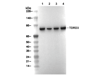 Lane 1: SH-SY5Y, Lane 2: 3T3, Lane 3: H-4-II-E, Lane 4: COS-7