
|
How to Cite 1. For In-Text Citation (Materials & Methods): 2. For Key Resources Table: |
||
|
Toll Free: (877) 796-6397 -- USA and Canada only -- |
Fax: +1-832-582-8590 Orders: +1-832-582-8158 |
Tech Support: +1-832-582-8158 Ext:3 Please provide your Order Number in the email. We strive to reply to |
Biological Description
| Specificity | TAZ/YAP Antibody (Rabbit mAb) [B1F12] detects endogenous levels of total TAZ and YAP proteins. |
|---|---|
| Background | YAP and TAZ are homologous transcriptional co‑activators that function as central effectors of the Hippo signaling cascade and as integrators of mechanical, morphogen and metabolic cues, translating changes in cell density, polarity, matrix stiffness and growth factor context into gene expression programs that control organ growth, progenitor cell amplification, tissue homeostasis and tumor behavior. Structurally, both proteins lack an intrinsic DNA‑binding domain and instead carry one or more WW domains that recognize PPxY motifs in partner transcription factors, a coiled‑coil region and a C‑terminal PDZ‑binding motif that supports interactions with TEAD1–4 and other nuclear partners; YAP also contains an SH3‑binding motif and multiple serine and threonine phosphorylation sites, including canonical Hippo sites such as human Ser127 (mouse Ser112), that govern its localization and stability. In the canonical Hippo pathway, upstream MST1/2–SAV1 and MAP4Ks activate LATS1/2–MOB1 complexes, which directly phosphorylate YAP and TAZ on multiple serine residues; this phosphorylation creates 14‑3‑3 binding sites that cause nuclear exclusion and cytoplasmic sequestration and primes adjacent phosphodegrons for secondary phosphorylation by casein kinase 1δ/ε, leading to SCFβ‑TrCP‑mediated ubiquitination and proteasomal degradation, so that activation of the kinase module concludes in inhibition of YAP/TAZ‑dependent transcription. When Hippo signaling is low, YAP/TAZ accumulate in the nucleus and bind TEAD transcription factors and other partners such as AP‑1 at distal enhancers and some promoters, where they modulate transcriptional pause release, H3K27 acetylation and nucleosome occupancy to drive expression of genes involved in cell cycle progression, survival, stemness and epithelial–mesenchymal transition, including CTGF, CYR61, AREG, BCL‑2 and other regulators of growth and anti‑apoptotic signaling. YAP and TAZ also act as primary sensors of the mechanical and architectural state of tissues: their activity reflects cell shape, adhesion and cytoskeletal tension, and they are regulated by Rho GTPases, GPCRs, mevalonate pathway activity and Wnt signals, so that changes in matrix stiffness, cell crowding or junctional integrity alter Hippo kinase activity and directly impinge on YAP/TAZ nuclear localization and transcriptional output. In stem and progenitor cell biology, YAP/TAZ sustain amplification of tissue‑specific progenitors during renewal and regeneration and influence lineage decisions in a context‑dependent manner; in mesenchymal stem cells, TAZ interacts with Runx2 and PPARγ to bias differentiation between osteogenic and adipogenic fates, and more broadly, YAP/TAZ complexes with TEAD control survival and self‑renewal of neural, intestinal, liver and skin stem cell populations. In cancer, YAP/TAZ can reprogram differentiated tumor cells into cancer stem‑like cells, promote metastasis and confer chemoresistance by upregulating pro‑survival and pro‑migration genes and by reshaping the tumor microenvironment: YAP/TAZ activation in tumor and stromal cells enhances fibrosis and angiogenesis, modulates immune checkpoints and cytokine production and alters innate immune cell behavior, collectively facilitating immune evasion and treatment resistance. At the level of immune and inflammatory signaling, YAP/TAZ regulate macrophage and other innate immune cell functions, where dysregulated YAP/TAZ activity hampers tissue repair and contributes to chronic inflammation and impaired regeneration, linking mechanochemical inputs from tissue remodeling to immune cell transcriptional programs. YAP also shows context‑dependent roles in apoptosis: in many cancers YAP/TAZ reduce apoptosis by increasing BCL‑2 and decreasing BAX, while under DNA damage conditions YAP can be phosphorylated by c‑Abl on Tyr357 and switch partners from TEAD to p73, driving pro‑apoptotic gene expression and cell death, illustrating that YAP’s impact on survival depends on upstream signaling state and binding partner repertoire. |
Usage Information
| Application | WB, IP, IHC | Dilution |
|
||||||
|---|---|---|---|---|---|---|---|---|---|
| Reactivity | Human, Mouse, Rat, Monkey | ||||||||
| Source | Rabbit Monoclonal Antibody | MW | 44 kDa | ||||||
| Storage Buffer | PBS, pH 7.2+50% Glycerol+0.05% BSA+0.01% NaN3 | Storage (from the date of receipt) |
-20°C (avoid freeze-thaw cycles), 2 years | ||||||
| WB |
Experimental Protocol:
Sample preparation
1. Tissue: Lyse the tissue sample by adding an appropriate volume of ice-cold RIPA/NP-40 Lysis Buffer (containing Protease Inhibitor Cocktail),and homogenize the tissue at a low temperature or lyse it by sonication on ice, then incubate on ice for 30 minutes. 2. Adherent cell: Aspirate the culture medium and wash the cells with ice-cold PBS twice. Lyse the cells by adding an appropriate volume of RIPA/NP-40 Lysis Buffer (containing Protease Inhibitor Cocktail) , sonicate to lyse the cells, and incubate on ice for 30 minutes. 3. Suspension cell: Transfer the culture medium to a pre-cooled centrifuge tube. Centrifuge and aspirate the supernatant. Wash the cells with ice-cold PBS twice. Lyse the cells by adding an appropriate volume of RIPA/NP-40 Lysis Buffer (containing Protease Inhibitor Cocktail) , sonicate to lyse the cells, and incubate on ice for 30 minutes. 4. Place the lysate into a pre-cooled microcentrifuge tube. Centrifuge at 4°C for 15 min. Collect the supernatant;
5. Remove a small volume of lysate to determine the protein concentration;
6. Combine the lysate with protein loading buffer. Boil 20 µL sample under 95-100°C for 5 min. Centrifuge for 5 min after cool down on ice.
Electrophoretic separation
1. According to the concentration of extracted protein, load appropriate amount of protein sample and marker onto SDS-PAGE gels for electrophoresis. Recommended separating gel (lower gel) concentration: 10%. Reference Table for Selecting SDS-PAGE Separation Gel Concentrations 2. Power up 80V for 30 minutes. Then the power supply is adjusted (110 V~150 V), the Marker is observed, and the electrophoresis can be stopped when the indicator band of the predyed protein Marker where the protein is located is properly separated. (Note that the current should not be too large when electrophoresis, too large current (more than 150 mA) will cause the temperature to rise, affecting the result of running glue. If high currents cannot be avoided, an ice bath can be used to cool the bath.)
Transfer membrane
1. Take out the converter, soak the clip and consumables in the pre-cooled converter;
2. Activate PVDF membrane with methanol for 1 min and rinse with transfer buffer;
3. Install it in the order of "black edge of clip - sponge - filter paper - filter paper - glue -PVDF membrane - filter paper - filter paper - sponge - white edge of clip"; 4. The protein was electrotransferred to PVDF membrane. ( 0.45 µm PVDF membrane is recommended ) Reference Table for Selecting PVDF Membrane Pore Size Specifications Recommended conditions for wet transfer: 200 mA, 120 min. ( Note that the transfer conditions can be adjusted according to the protein size. For high-molecular-weight proteins, a higher current and longer transfer time are recommended. However, ensure that the transfer tank remains at a low temperature to prevent gel melting.)
Block
1. After electrotransfer, wash the film with TBST at room temperature for 5 minutes;
2. Incubate the film in the blocking solution for 1 hour at room temperature;
3. Wash the film with TBST for 3 times, 5 minutes each time.
Antibody incubation
1. Use primary antibody dilution buffer to prepare the primary antibody working liquid (recommended dilution ratio for primary antibody 1:1000), gently shake and incubate with the film at 4°C overnight; 2. Wash the film with TBST 3 times, 5 minutes each time;
3. Add the secondary antibody to the blocking solution and incubate with the film gently at room temperature for 1 hour;
4. After incubation, wash the film with TBST 3 times for 5 minutes each time.
Antibody staining
1. Add the prepared ECL luminescent substrate (or select other color developing substrate according to the second antibody) and mix evenly;
2. Incubate with the film for 1 minute, remove excess substrate (keep the film moist), wrap with plastic film, and expose in the imaging system.
|
| IHC |
Experimental Protocol:
Deparaffinization/Rehydration
1. Deparaffinize/hydrate sections:
2. Incubate sections in three washes of xylene for 5 min each.
3. Incubate sections in two washes of 100% ethanol for 10 min each.
4. Incubate sections in two washes of 95% ethanol for 10 min each.
5. Wash sections two times in dH2O for 5 min each.
6.Antigen retrieval: For Citrate: Heat slides in a microwave submersed in 1X citrate unmasking solution until boiling is initiated; continue with 10 min at a sub-boiling temperature (95°-98°C). Cool slides on bench top for 30 min.
Staining
1. Wash sections in dH2O three times for 5 min each.
2. Incubate sections in 3% hydrogen peroxide for 10 min.
3. Wash sections in dH2O two times for 5 min each.
4. Wash sections in wash buffer for 5 min.
5. Block each section with 100–400 µl of blocking solution for 1 hr at room temperature.
6. Remove blocking solution and add 100–400 µl primary antibody diluent in to each section. Incubate overnight at 4°C.
7. Remove antibody solution and wash sections with wash buffer three times for 5 min each.
8. Cover section with 1–3 drops HRPas needed. Incubate in a humidified chamber for 30 min at room temperature.
9. Wash sections three times with wash buffer for 5 min each.
10. Add DAB Chromogen Concentrate to DAB Diluent and mix well before use.
11. Apply 100–400 µl DAB to each section and monitor closely. 1–10 min generally provides an acceptable staining intensity.
12. Immerse slides in dH2O.
13. If desired, counterstain sections with hematoxylin.
14. Wash sections in dH2O two times for 5 min each.
15. Dehydrate sections: Incubate sections in 95% ethanol two times for 10 sec each; Repeat in 100% ethanol, incubating sections two times for 10 sec each; Repeat in xylene, incubating sections two times for 10 sec each.
16. Mount sections with coverslips and mounting medium.
|
References
|
Application Data
WB
Validated by Selleck
-
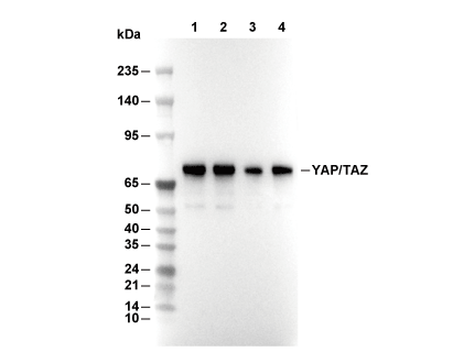 Lane 1: 293T, Lane 2: PANC-1, Lane 3: Hela, Lane 4: A204