|
How to Cite 1. For In-Text Citation (Materials & Methods): 2. For Key Resources Table: |
||
|
Toll Free: (877) 796-6397 -- USA and Canada only -- |
Fax: +1-832-582-8590 Orders: +1-832-582-8158 |
Tech Support: +1-832-582-8158 Ext:3 Please provide your Order Number in the email. We strive to reply to |
Biological Description
| Specificity | TAX1BP1 Antibody [B22F23] detects endogenous levels of total TAX1BP1 protein. |
|---|---|
| Background | TAX1BP1 (Tax1-binding protein 1), a modular adaptor of ~789 amino acids initially identified as an HTLV-1 Tax interactor, features an N-terminal SKICH domain for binding NAP1/RB1CC1 in autophagy initiation, a central coiled-coil/helix-loop-helix region promoting homodimerization, a 14-3-3 motif for phosphoregulation, and C-terminal tandem zinc finger (UBZ1/2) domains that specifically recognize K63-linked ubiquitin chains on substrates like TRAF6, RIP1, and TBK1. In its core negative feedback role within innate immunity, TAX1BP1 acts as a ubiquitin-sensing scaffold that rapidly recruits the deubiquitinase A20 (TNFAIP3) to polyubiquitinated signaling hubs in TNFR/IL-1R complexes upon ligand stimulation, IKKα phosphorylates TAX1BP1 at Ser593/Ser624 to trigger this assembly, enabling A20's OTU domain to cleave K63-ubiquitin while deploying its ZnF domain to add K48-chains for proteasomal degradation of TRAF6/RIPK1, thereby terminating canonical NF-κB and IRF3 activation to prevent excessive inflammation; dual UBZ domains bind diubiquitin with high affinity via conserved arginines engaging the proximal ubiquitin's Ile44 patch. TAX1BP1 drives selective autophagy by linking ubiquitinated cargoes (aggregates, bacteria, mitochondria) to the nascent phagophore, SKICH docks RB1CC1/FIP200 for ULK1 complex recruitment and NAP1/TBK1 for phosphorylation maturation, while UBZs capture substrates and C-terminal motifs bind LC3/GABARAP for membrane enclosure, facilitating xenophagy, mitophagy, and aggrephagy; Myosin VI interaction aids autophagosome-endosome fusion. Dysregulation promotes autoimmunity (e.g., lupus via sustained NF-κB), chronic inflammation, neurodegeneration (impaired protein clearance), tumorigenesis (uncontrolled IRF3/NF-κB), and viral persistence (e.g., HTLV-1 Tax evasion), with cytoplasmic localization shifting to the nucleus under stress. |
Usage Information
| Application | WB, IP | Dilution |
|
||||
|---|---|---|---|---|---|---|---|
| Reactivity | Human | ||||||
| Source | Rabbit Monoclonal Antibody | MW | 92 kDa | ||||
| Storage Buffer | PBS, pH 7.2+50% Glycerol+0.05% BSA+0.01% NaN3 | Storage (from the date of receipt) |
-20°C (avoid freeze-thaw cycles), 2 years | ||||
| WB |
Experimental Protocol:
Sample preparation
1. Tissue: Lyse the tissue sample by adding an appropriate volume of ice-cold RIPA/NP-40 Lysis Buffer (containing Protease Inhibitor Cocktail),and homogenize the tissue at a low temperature. 2. Adherent cell: Aspirate the culture medium and wash the cells with ice-cold PBS twice. Lyse the cells by adding an appropriate volume of RIPA/NP-40 Lysis Buffer (containing Protease Inhibitor Cocktail) and put the sample on ice for 5 min. 3. Suspension cell: Transfer the culture medium to a pre-cooled centrifuge tube. Centrifuge and aspirate the supernatant. Wash the cells with ice-cold PBS twice. Lyse the cells by adding an appropriate volume of RIPA/NP-40 Lysis Buffer (containing Protease Inhibitor Cocktail) and put the sample on ice for 5 min. 4. Place the lysate into a pre-cooled microcentrifuge tube. Centrifuge at 4°C for 15 min. Collect the supernatant;
5. Remove a small volume of lysate to determine the protein concentration;
6. Combine the lysate with protein loading buffer. Boil 20 µL sample under 95-100°C for 5 min. Centrifuge for 5 min after cool down on ice.
Electrophoretic separation
1. According to the concentration of extracted protein, load appropriate amount of protein sample and marker onto SDS-PAGE gels for electrophoresis. Recommended separating gel (lower gel) concentration: 10%. Reference Table for Selecting SDS-PAGE Separation Gel Concentrations 2. Power up 80V for 30 minutes. Then the power supply is adjusted (110 V~150 V), the Marker is observed, and the electrophoresis can be stopped when the indicator band of the predyed protein Marker where the protein is located is properly separated. (Note that the current should not be too large when electrophoresis, too large current (more than 150 mA) will cause the temperature to rise, affecting the result of running glue. If high currents cannot be avoided, an ice bath can be used to cool the bath.)
Transfer membrane
1. Take out the converter, soak the clip and consumables in the pre-cooled converter;
2. Activate PVDF membrane with methanol for 1 min and rinse with transfer buffer;
3. Install it in the order of "black edge of clip - sponge - filter paper - filter paper - glue -PVDF membrane - filter paper - filter paper - sponge - white edge of clip"; 4. The protein was electrotransferred to PVDF membrane. ( 0.45 µm PVDF membrane is recommended ) Reference Table for Selecting PVDF Membrane Pore Size Specifications Recommended conditions for wet transfer: 200 mA, 120 min. ( Note that the transfer conditions can be adjusted according to the protein size. For high-molecular-weight proteins, a higher current and longer transfer time are recommended. However, ensure that the transfer tank remains at a low temperature to prevent gel melting.)
Block
1. After electrotransfer, wash the film with TBST at room temperature for 5 minutes;
2. Incubate the film in the blocking solution for 1 hour at room temperature;
3. Wash the film with TBST for 3 times, 5 minutes each time.
Antibody incubation
1. Use 5% skim milk powder to prepare the primary antibody working liquid (recommended dilution ratio for primary antibody 1:1000), gently shake and incubate with the film at 4°C overnight; 2. Wash the film with TBST 3 times, 5 minutes each time;
3. Add the secondary antibody to the blocking solution and incubate with the film gently at room temperature for 1 hour;
4. After incubation, wash the film with TBST 3 times for 5 minutes each time.
Antibody staining
1. Add the prepared ECL luminescent substrate (or select other color developing substrate according to the second antibody) and mix evenly;
2. Incubate with the film for 1 minute, remove excess substrate (keep the film moist), wrap with plastic film, and expose in the imaging system.
|
References
|
Application Data
WB
Validated by Selleck
-
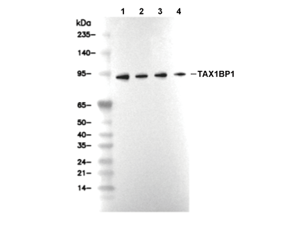 Lane 1: NCI-H226, Lane 2: U-937, Lane 3: Hela, Lane 4: HepG2