
|
How to Cite 1. For In-Text Citation (Materials & Methods): 2. For Key Resources Table: |
||
|
Toll Free: (877) 796-6397 -- USA and Canada only -- |
Fax: +1-832-582-8590 Orders: +1-832-582-8158 |
Tech Support: +1-832-582-8158 Ext:3 Please provide your Order Number in the email. We strive to reply to |
Biological Description
| Specificity | Snail + Slug N-terminal Antibody (Mouse mAb) [E10J13] detects endogenous levels of total Snail and Slug proteins, typically through their N-terminal regions. |
|---|---|
| Background | SNAIL and SLUG, also designated SNAI1 and SNAI2, belong to the SNAIL family of zinc finger transcription factors that drive epithelial-mesenchymal transition, and both proteins share a conserved N-terminal SNAG domain that functions as the primary repressive module distinct from the C-terminal zinc finger DNA-binding region. This SNAG domain adopts a structure resembling the N-terminal tail of histone H3, allowing it to act as a pseudosubstrate hook that docks directly into the amino oxidase domain of the histone lysine-specific demethylase LSD1, and this interaction stabilizes a ternary Snail1-LSD1-CoREST complex at target gene promoters, most notably the E-cadherin promoter, CDH1. Once this complex is assembled, transcriptional silencing proceeds through a defined multistage sequence: HDAC1 and HDAC2 within the CoREST-Snail1-LSD1 complex first deacetylate histone H3 and H4, Polycomb repressive complex 2 is then recruited in a manner that requires both an intact Snail1-binding site on the promoter and the SNAG domain itself, directing trimethylation of histone H3 lysine 27, and LSD1 subsequently removes the activating H3K4 methylation mark, together converting an active promoter into a stably silenced one. In Slug, the SNAG domain, spanning the extreme N-terminus, recruits the corepressor NCoR directly, while a separate central region termed the SLUG domain recruits CtBP1, and functional dissection using domain-deletion constructs shows that the SNAG domain is absolutely required for EMT induction, whereas the SLUG domain contributes to efficient E-cadherin repression but exerts a negative modulatory effect on SNAG-driven EMT induction and on NCoR corepressor activity when deleted. Both SNAIL and SLUG require their SNAG domains for E-cadherin promoter repression, but the domain composition governing corepressor recruitment differs between the two paralogs, with Slug relying on two distinct, functionally separable domains that recruit different corepressor complexes rather than a single shared repressive interface. This SNAG-LSD1 interaction is directly druggable: pharmacological inhibition of LSD1 with the enzymatic inhibitor Parnate, or competitive blockade using a cell-permeable TAT-SNAG peptide corresponding to the Slug SNAG domain, disrupts Slug-dependent repression of the E-cadherin promoter and suppresses motility and invasion across tumor cell lines of varying origin without affecting cell proliferation, demonstrating that the SNAG domain interaction with chromatin-modifying enzymes is mechanistically separable from other SNAIL and SLUG functions and represents a defined point of therapeutic intervention in EMT-driven cancer invasion. |
Usage Information
| Application | IHC, IF | Dilution |
|
||||
|---|---|---|---|---|---|---|---|
| Reactivity | Human | ||||||
| Source | Mouse Monoclonal Antibody | MW | 29 kDa | ||||
| Storage Buffer | PBS, pH 7.2+50% Glycerol+0.05% BSA+0.01% NaN3 | Storage (from the date of receipt) |
-20°C (avoid freeze-thaw cycles), 2 years | ||||
| IHC |
Experimental Protocol:
Deparaffinization/Rehydration
1. Deparaffinize/hydrate sections:
2. Incubate sections in three washes of xylene for 5 min each.
3. Incubate sections in two washes of 100% ethanol for 10 min each.
4. Incubate sections in two washes of 95% ethanol for 10 min each.
5. Wash sections two times in dH2O for 5 min each.
6.Antigen retrieval: For Citrate: Heat slides in a microwave submersed in 1X citrate unmasking solution until boiling is initiated; continue with 10 min at a sub-boiling temperature (95°-98°C). Cool slides on bench top for 30 min.
Staining
1. Wash sections in dH2O three times for 5 min each.
2. Incubate sections in 3% hydrogen peroxide for 10 min.
3. Wash sections in dH2O two times for 5 min each.
4. Wash sections in wash buffer for 5 min.
5. Block each section with 100–400 µl of blocking solution for 1 hr at room temperature.
6. Remove blocking solution and add 100–400 µl primary antibody diluent in to each section. Incubate overnight at 4°C.
7. Remove antibody solution and wash sections with wash buffer three times for 5 min each.
8. Cover section with 1–3 drops HRPas needed. Incubate in a humidified chamber for 30 min at room temperature.
9. Wash sections three times with wash buffer for 5 min each.
10. Add DAB Chromogen Concentrate to DAB Diluent and mix well before use.
11. Apply 100–400 µl DAB to each section and monitor closely. 1–10 min generally provides an acceptable staining intensity.
12. Immerse slides in dH2O.
13. If desired, counterstain sections with hematoxylin.
14. Wash sections in dH2O two times for 5 min each.
15. Dehydrate sections: Incubate sections in 95% ethanol two times for 10 sec each; Repeat in 100% ethanol, incubating sections two times for 10 sec each; Repeat in xylene, incubating sections two times for 10 sec each.
16. Mount sections with coverslips and mounting medium.
|
| IF |
Experimental Protocol:
Sample Preparation
1. Adherent Cells: Place a clean, sterile coverslip in a culture dish. Once the cells grow to near confluence as a monolayer, remove the coverslip for further use.
2. Suspension Cells: Seed the cells onto a clean, sterile slide coated with poly-L-lysine.
3. Frozen Sections: Allow the slide to thaw at room temperature. Wash it with pure water or PBS for 2 times, 3 minutes each time.
4. Paraffin Sections: Deparaffinization and rehydration. Wash the slide with pure water or PBS for 3 times, 3 minutes each time. Then perform antigen retrieval.
Fixation
1. Fix the cell coverslips/spots or tissue sections at room temperature using a fixative such as 4% paraformaldehyde (4% PFA) for 10-15 minutes.
2. Wash the sample with PBS for 3 times, 3 minutes each time.
Permeabilization
1.Add a detergent such as 0.1–0.3% Triton X-100 to the sample and incubate at room temperature for 10–20 minutes.
(Note: This step is only required for intracellular antigens. For antigens expressed on the cell membrane, this step is unnecessary.)
Wash the sample with PBS for 3 times, 3 minutes each time.
Blocking
Add blocking solution and incubate at room temperature for at least 1 hour. (Common blocking solutions include: serum from the same source as the secondary antibody, BSA, or goat serum.)
Note: Ensure the sample remains moist during and after the blocking step to prevent drying, which can lead to high background.
Immunofluorescence Staining (Day 1)
1. Remove the blocking solution and add the diluted primary antibody.
2. Incubate the sample in a humidified chamber at 4°C overnight.
Immunofluorescence Staining (Day 2)
1. Remove the primary antibody and wash with PBST for 3 times, 5 minutes each time.
2. Add the diluted fluorescent secondary antibody and incubate in the dark at 4°C for 1–2 hours.
3. Remove the secondary antibody and wash with PBST for 3 times, 5 minutes each time.
4. Add diluted DAPI and incubate at room temperature in the dark for 5–10 minutes.
5. Wash with PBST for 3 times, 5 minutes each time.
Mounting
1. Mount the sample with an anti-fade mounting medium.
2. Allow the slide to dry at room temperature overnight in the dark.
3. Store the slide in a slide storage box at 4°C, protected from light.
|
References
|
Application Data
IHC
Validated by Selleck
-
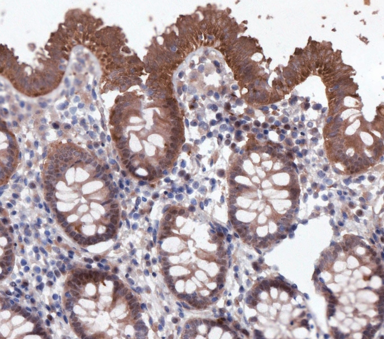 Immunohistochemical analysis of formalin fixed paraffin embedded human colon tissue with F3674 at 1:5000 dilution.