|
How to Cite 1. For In-Text Citation (Materials & Methods): 2. For Key Resources Table: |
||
|
Toll Free: (877) 796-6397 -- USA and Canada only -- |
Fax: +1-832-582-8590 Orders: +1-832-582-8158 |
Tech Support: +1-832-582-8158 Ext:3 Please provide your Order Number in the email. We strive to reply to |
Biological Description
| Specificity | SCXA Antibody [M20P22] detects endogenous levels of total SCXA protein. |
|---|---|
| Background | Scleraxis A (SCXA) is a basic helix–loop–helix transcription factor of the scleraxis family that marks tendon and ligament lineages and functions as a central regulator of tenogenic differentiation and tendon connective tissue organization throughout development and into postnatal life. The protein contains a single basic helix–loop–helix DNA‑binding and dimerization domain that allows homodimer or heterodimer formation with other bHLH factors and binding to E‑box motifs in regulatory regions of extracellular matrix and cytoskeletal genes associated with tendon identity, positioning SCXA within transcriptional complexes that activate tendon‑specific programs while constraining alternative mesenchymal fates. SCXA expression defines early tendon progenitors in limb and axial mesenchyme, is maintained in differentiating tenocytes, and shows selective enrichment in force‑transmitting and intermuscular tendons, where it coordinates the transition from progenitor pools to elongated, matrix‑producing tendon cells and supports assembly of dense, longitudinally aligned collagen fibrils required for efficient muscle‑to‑bone force transmission. SCXA directly regulates a broad set of genes involved in extracellular matrix synthesis and remodeling, including fibrillar collagens, small leucine‑rich proteoglycans, cell–matrix adhesion components, and regulators of actin organization and cell shape, and controls multiple cellular processes such as proliferation, cytoskeletal dynamics, and matrix deposition that together establish tendon mechanical properties and structural integrity. SCXA activity integrates with growth factor pathways such as TGF‑β signaling, which maintains tendon cell fate and supports continued expression of tendon differentiation markers, and with positional cues in developing limbs that distinguish force‑transmitting tendons from muscle‑anchoring tendons, thereby linking extracellular signaling environments to stable tenocyte gene expression states. Disruption of SCXA function leads to severe defects in the differentiation and organization of force‑transmitting tendons with reduced, disorganized tendon matrix and impaired tendon‑to‑bone attachment (enthesis) structure, while some anchoring tendons remain comparatively preserved. |
Usage Information
| Application | IHC | Dilution |
|
||
|---|---|---|---|---|---|
| Reactivity | Rat, Mouse | ||||
| Source | Rabbit Monoclonal Antibody | MW | 22 kDa | ||
| Storage Buffer | PBS, pH 7.2+50% Glycerol+0.05% BSA+0.01% NaN3 | Storage (from the date of receipt) |
-20°C (avoid freeze-thaw cycles), 2 years | ||
References
|
Application Data
IHC
Validated by Selleck
-
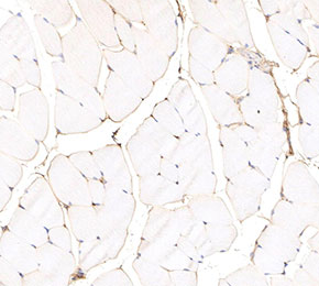 Immunohistochemical analysis of formalin fixed paraffin embedded mouse tendons tissue with F3991 at 1:500 dilution.