|
Toll Free: (877) 796-6397 -- USA and Canada only -- |
Fax: +1-832-582-8590 Orders: +1-832-582-8158 |
Tech Support: +1-832-582-8158 Ext:3 Please provide your Order Number in the email. |
Biological Description
| Specificity | PLD2 Rabbit mAb recognizes endogenous levels of total PLD2 protein. |
|---|---|
| Background | Phosphatidylcholine-specific phospholipase D (PLD) catalyzes the hydrolysis of phosphatidylcholine (PC) to generate choline and phosphatidic acid (PA), with PA serving as a precursor to the second messenger diacylglycerol (DAG). PLD exists in two main isoforms: PLD1 and PLD2. PLD2 is primarily localized at the plasma membrane under basal conditions, where it associates with receptors in lipid rafts, whereas PLD1 is found in perinuclear membranes and translocates to the plasma membrane upon cell stimulation. Activation of PLD2 is mediated by protein kinase C (PKC) and receptor (e.g., EGFR, PDGFR) and non-receptor tyrosine kinases (e.g., Src, JAK3), whereas PLD1 is activated by PKC, casein kinase-II, and small GTPases such as ARF and RHO. PLD2 exhibits higher basal activity compared to PLD1 and functions not only as a phospholipase but also as a guanine nucleotide exchange factor (GEF). PLD2 activity is modulated through complex phosphorylation and dephosphorylation pathways, primarily on tyrosine residues, involving interactions with proteins like S6K, Grb2, Sos, WASp, and Rac2. The PLD2 isoform is highly expressed in various cancers, including colorectal and breast cancers, and also acts as a GEF for the small GTPase Rac2, independent of its phospholipase function. |
Usage Information
| Application | WB | Dilution |
|
||
|---|---|---|---|---|---|
| Reactivity | Human | ||||
| Source | Rabbit | MW | 95 kDa | ||
| Storage Buffer | PBS, pH 7.2+50% Glycerol+0.05% BSA+0.01% NaN₃ | Storage (from the date of receipt) |
–20°C (avoid freeze-thaw cycles), 2 years | ||
| WB |
Experimental Protocol:
Sample preparation
1. Tissue: Lyse the tissue sample by adding an appropriate volume of ice-cold RIPA/NP-40 Lysis Buffer (containing Protease Inhibitor Cocktail),and homogenize the tissue at a low temperature or lyse it by sonication on ice, then incubate on ice for 30 minutes. 2. Adherent cell: Aspirate the culture medium and transfer the cells into an EP tube. Wash the cells with ice-cold PBS twice. Add an appropriate volume of RIPA/NP-40 Lysis Buffer (containing Protease Inhibitor Cocktail), sonicate to lyse the cells, and incubate on ice for 30 minutes. 3. Suspension cell: Transfer the culture medium to a pre-cooled centrifuge tube. Centrifuge and aspirate the supernatant. Wash the cells with ice-cold PBS twice.Add an appropriate volume of RIPA/NP-40 Lysis Buffer (containing Protease Inhibitor Cocktail), sonicate to lyse the cells, and incubate on ice for 30 minutes. 4. Place the lysate into a pre-cooled microcentrifuge tube. Centrifuge at 4°C for 15 min. Collect the supernatant;
5. Remove a small volume of lysate to determine the protein concentration;
6. Combine the lysate with protein loading buffer. Boil 20 µL sample under 95-100°C for 5 min. Centrifuge for 5 min after cool down on ice.
Electrophoretic separation
1. According to the concentration of extracted protein, load appropriate amount of protein sample and marker onto SDS-PAGE gels for electrophoresis. Recommended separating gel (lower gel) concentration: 10%. Reference Table for Selecting SDS-PAGE Separation Gel Concentrations 2. Power up 80V for 30 minutes. Then the power supply is adjusted (110 V~150 V), the Marker is observed, and the electrophoresis can be stopped when the indicator band of the predyed protein Marker where the protein is located is properly separated. (Note that the current should not be too large when electrophoresis, too large current (more than 150 mA) will cause the temperature to rise, affecting the result of running glue. If high currents cannot be avoided, an ice bath can be used to cool the bath.)
Transfer membrane
1. Take out the converter, soak the clip and consumables in the pre-cooled converter;
2. Activate PVDF membrane with methanol for 1 min and rinse with transfer buffer;
3. Install it in the order of "black edge of clip - sponge - filter paper - filter paper - glue -PVDF membrane - filter paper - filter paper - sponge - white edge of clip"; 4. The protein was electrotransferred to PVDF membrane. ( 0.45 µm PVDF membrane is recommended ) Reference Table for Selecting PVDF Membrane Pore Size Specifications Recommended conditions for wet transfer: 200 mA, 120 min. ( Note that the transfer conditions can be adjusted according to the protein size. For high-molecular-weight proteins, a higher current and longer transfer time are recommended. However, ensure that the transfer tank remains at a low temperature to prevent gel melting.)
Block
1. After electrotransfer, wash the film with TBST at room temperature for 5 minutes;
2. Incubate the film in the blocking solution for 1 hour at room temperature;
3. Wash the film with TBST for 3 times, 5 minutes each time.
Antibody incubation
1. Use 5% skim milk powder to prepare the primary antibody working liquid (recommended dilution ratio for primary antibody 1:1000), gently shake and incubate with the film at 4°C overnight; 2. Wash the film with TBST 3 times, 5 minutes each time;
3. Add the secondary antibody to the blocking solution and incubate with the film gently at room temperature for 1 hour;
4. After incubation, wash the film with TBST 3 times for 5 minutes each time.
Antibody staining
818. Add the prepared ECL luminescent substrate (or select other color developing substrate according to the second antibody) and mix evenly;
2. Incubate with the film for 1 minute, remove excess substrate (keep the film moist), wrap with plastic film, and expose in the imaging system.
|
Application Data
WB
Validated by Selleck
-
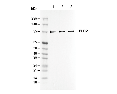 Lane 1: Hela
Lane 2: A549
Lane 3: HUVEC
IHC
Validated by Selleck
-
 Immunohistochemical analysis of formalin fixed paraffin embedded human Colorectal cancer tissue with F1493 at 1/100 dilution.