|
How to Cite 1. For In-Text Citation (Materials & Methods): 2. For Key Resources Table: |
||
|
Toll Free: (877) 796-6397 -- USA and Canada only -- |
Fax: +1-832-582-8590 Orders: +1-832-582-8158 |
Tech Support: +1-832-582-8158 Ext:3 Please provide your Order Number in the email. We strive to reply to |
Biological Description
| Specificity | PAR2 Antibody [J17G12] detects endogenous levels of total PAR2 protein. |
|---|---|
| Background | PAR2 belongs to the protease-activated receptor family of G protein-coupled receptors alongside PAR1, PAR3, and PAR4, distinguished by proteolytic unmasking of cryptic N-terminal tethered ligands rather than diffusible agonists. PAR2 organizes seven transmembrane helices with an extracellular N-terminus featuring the SFLLRN sequence that, upon cleavage at Arg36 by trypsin-like serine proteases or mast cell tryptase, folds back to engage the second extracellular loop triggering G protein dissociation. Protease activation induces conformational shift where TM6 outward movement exposes DRY motif for Gαq/11 coupling that stimulates phospholipase Cβ hydrolysis of PIP2 into IP3 and DAG, mobilizing intracellular calcium for smooth muscle contraction alongside PKC-mediated Raf-MEK-ERK cascade activation driving cytokine transcription via Elk-1 and NF-κB nuclear translocation. β-arrestin-2 recruitment sustains MAPK signaling while promoting clathrin-mediated endocytosis that either recycles functional receptors or targets them for lysosomal degradation, with biased agonism from neutrophil elastase cleavage at distinct sites yielding G12/13-RhoA outputs favoring migration over secretion. Canonical trypsin cleavage at R36-S37 activates comprehensive Gq/11, Gi, and G12/13 pathways, while non-canonical sites produce signaling bias through differential β-arrestin engagement. PAR2 governs epithelial barrier restitution through Rac1-dependent lamellipodia protrusion and sensory neuron hyperexcitability via TRPV1 sensitization during inflammation, with highest expression in colon enterocytes, airway epithelium, and dorsal root ganglia reflecting roles in mucosal defense and pain transmission. Signal peptide shielding prevents premature intracellular activation during biosynthesis, ensuring surface competence. Constitutive PAR2 upregulation drives colitis-associated tumorigenesis and arthritic synovitis through IL-8 and MMP9 induction. |
Usage Information
| Application | WB | Dilution |
|
||
|---|---|---|---|---|---|
| Reactivity | Human | ||||
| Source | Rabbit Monoclonal Antibody | MW | 32 kDa | ||
| Storage Buffer | PBS, pH 7.2+50% Glycerol+0.05% BSA+0.01% NaN3 | Storage (from the date of receipt) |
-20°C (avoid freeze-thaw cycles), 2 years | ||
References
|
Application Data
WB
Validated by Selleck
-
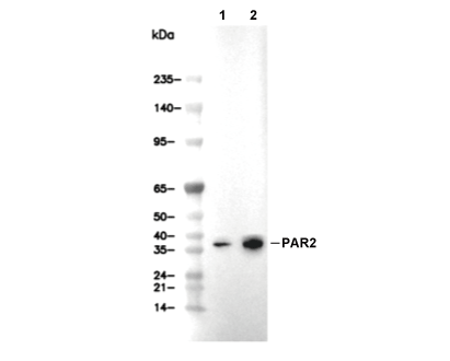 Lane 1: HCT116, Lane 2: HepG2