|
How to Cite 1. For In-Text Citation (Materials & Methods): 2. For Key Resources Table: |
||
|
Toll Free: (877) 796-6397 -- USA and Canada only -- |
Fax: +1-832-582-8590 Orders: +1-832-582-8158 |
Tech Support: +1-832-582-8158 Ext:3 Please provide your Order Number in the email. We strive to reply to |
Biological Description
| Specificity | Nicastrin Antibody [D2C5] detects endogenous levels of total Nicastrin protein. |
|---|---|
| Background | Nicastrin functions as the essential glycoprotein subunit of the γ-secretase protease complex alongside presenilin, APH-1, and PEN-2, enabling intramembrane proteolysis of type I transmembrane substrates following ectodomain shedding. Nicastrin organizes a large, heavily glycosylated extracellular domain with a substrate-binding pocket flanked by a single C-terminal transmembrane helix that anchors the complex within lipid rafts, while conserved glutamate and tyrosine residues in the DYIGS motif line a hydrophilic cleft recognizing free N-termini of cleaved substrates. The Nicastrin ectodomain directly captures the nascent amino terminus generated by α- or β-secretase cleavage of APP and Notch, recruiting these substrates into the active site through conformational stabilization that positions membrane stubs for presenilin-mediated endoproteolysis via two juxtaposed transmembrane aspartates. This docking mechanism exhibits substrate selectivity where mutations disrupting the binding pocket differentially impair Aβ40 versus Aβ42 production or Notch intracellular domain release, with Nicastrin stabilization preventing APH-1 and full-length presenilin degradation through subcomplex formation essential for Golgi-to-plasma membrane trafficking. Complex maturation requires sequential glycosylation of Nicastrin asparagines that shield hydrophobic surfaces during assembly, culminating in PEN-2-induced presenilin endoproteolysis that activates the catalytic core. Nicastrin coordinates neuronal differentiation through Notch signaling and synaptic plasticity via regulated intramembrane proteolysis of ErbB4 and cadherins, with ubiquitous expression ensuring a broad substrate repertoire across tissues. Dysregulation through familial mutations elevates Aβ42/40 ratios in Alzheimer's disease while disrupting developmental Notch patterning. |
Usage Information
| Application | WB | Dilution |
|
||
|---|---|---|---|---|---|
| Reactivity | Human, Mouse, Rat, Hamster, Monkey | ||||
| Source | Rabbit Monoclonal Antibody | MW | 110 kDa, 120 kDa | ||
| Storage Buffer | PBS, pH 7.2+50% Glycerol+0.05% BSA+0.01% NaN3 | Storage (from the date of receipt) |
-20°C (avoid freeze-thaw cycles), 2 years | ||
References
|
Application Data
WB
Validated by Selleck
-
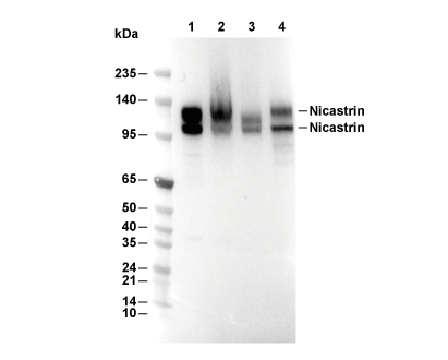 Lane 1: Hela, Lane 2: COS-7, Lane 3: C6, Lane 4: PC12