|
Toll Free: (877) 796-6397 -- USA and Canada only -- |
Fax: +1-832-582-8590 Orders: +1-832-582-8158 |
Tech Support: +1-832-582-8158 Ext:3 Please provide your Order Number in the email. |
Biological Description
| Specificity | Merlin Rabbit mAb recognizes endogenous levels of total Merlin protein. |
|---|---|
| Background | Merlin (also known as Schwannomin) is a tumor suppressor protein encoded by the NF2 gene and is a member of the ezrin/radixin/moesin (ERM) protein family. It is highly expressed in the nervous system and plays a crucial role in maintaining tissue architecture and preventing uncontrolled cell proliferation. Merlin's structure consists of an N-terminal FERM domain, a central α-helical region, and a C-terminal domain. The FERM domain is critical for protein-protein interactions and tumor suppressor activity, while the C-terminal domain interacts with the FERM domain to regulate Merlin's conformation and activity. Merlin functions by linking the cytoskeleton to the plasma membrane, regulating cell shape, migration, and growth. It acts as a negative regulator of the Hippo pathway, which controls cell growth and apoptosis. Merlin also inhibits Ras and Rac signaling pathways, contributing to contact-dependent growth inhibition. Merlin's activity is regulated by post-translational modifications, particularly phosphorylation. Phosphorylation at serine 518 by PAK2 or PKA promotes a closed, inactive conformation, reducing its tumor suppressor activity. Conversely, dephosphorylation enhances Merlin's tumor suppressor functions. Merlin can form dimers via FERM-FERM interactions, crucial for its interactions with binding partners and tumor suppressor activity. Merlin is also involved in regulating cell adhesion, particularly at cell-cell junctions, and plays a role in mechanotransduction. It interacts with CD44, NHERF1, and Angiomotin, to mediate its functions. Additionally, Merlin has been implicated in regulating fatty acid synthesis, expanding its role in cellular metabolism. Dysfunction of Merlin leads to the development of tumors, particularly schwannomas and meningiomas, characteristic of Neurofibromatosis Type 2. |
Usage Information
| Application | WB, IP, IF | Dilution |
|
||||||
|---|---|---|---|---|---|---|---|---|---|
| Reactivity | Human, Mouse, Rat, Monkey, Hamster | ||||||||
| Source | Rabbit | MW | 70 KDa | ||||||
| Storage Buffer | PBS, pH 7.2+50% Glycerol+0.05% BSA+0.01% NaN3 | Storage (from the date of receipt) |
-20°C (avoid freeze-thaw cycles), 2 years | ||||||
| WB |
Experimental Protocol:
Sample preparation
1. Tissue: Lyse the tissue sample by adding an appropriate volume of ice-cold RIPA/NP-40 Lysis Buffer (containing Protease Inhibitor Cocktail),and homogenize the tissue at a low temperature or lyse it by sonication on ice, then incubate on ice for 30 minutes. 2. Adherent cell: Aspirate the culture medium and transfer the cells into an EP tube. Wash the cells with ice-cold PBS twice. Add an appropriate volume of RIPA/NP-40 Lysis Buffer (containing Protease Inhibitor Cocktail), sonicate to lyse the cells, and incubate on ice for 30 minutes. 3. Suspension cell: Transfer the culture medium to a pre-cooled centrifuge tube. Centrifuge and aspirate the supernatant. Wash the cells with ice-cold PBS twice.Add an appropriate volume of RIPA/NP-40 Lysis Buffer (containing Protease Inhibitor Cocktail), sonicate to lyse the cells, and incubate on ice for 30 minutes. 4. Place the lysate into a pre-cooled microcentrifuge tube. Centrifuge at 4°C for 15 min. Collect the supernatant;
5. Remove a small volume of lysate to determine the protein concentration;
6. Combine the lysate with protein loading buffer. Boil 20 µL sample under 95-100°C for 5 min. Centrifuge for 5 min after cool down on ice.
Electrophoretic separation
1. According to the concentration of extracted protein, load appropriate amount of protein sample and marker onto SDS-PAGE gels for electrophoresis. Recommended separating gel (lower gel) concentration: 10%. Reference Table for Selecting SDS-PAGE Separation Gel Concentrations 2. Power up 80V for 30 minutes. Then the power supply is adjusted (110 V~150 V), the Marker is observed, and the electrophoresis can be stopped when the indicator band of the predyed protein Marker where the protein is located is properly separated. (Note that the current should not be too large when electrophoresis, too large current (more than 150 mA) will cause the temperature to rise, affecting the result of running glue. If high currents cannot be avoided, an ice bath can be used to cool the bath.)
Transfer membrane
1. Take out the converter, soak the clip and consumables in the pre-cooled converter;
2. Activate PVDF membrane with methanol for 1 min and rinse with transfer buffer;
3. Install it in the order of "black edge of clip - sponge - filter paper - filter paper - glue -PVDF membrane - filter paper - filter paper - sponge - white edge of clip"; 4. The protein was electrotransferred to PVDF membrane. ( 0.45 µm PVDF membrane is recommended ) Reference Table for Selecting PVDF Membrane Pore Size Specifications Recommended conditions for wet transfer: 200 mA, 120 min. ( Note that the transfer conditions can be adjusted according to the protein size. For high-molecular-weight proteins, a higher current and longer transfer time are recommended. However, ensure that the transfer tank remains at a low temperature to prevent gel melting.)
Block
1. After electrotransfer, wash the film with TBST at room temperature for 5 minutes;
2. Incubate the film in the blocking solution for 1 hour at room temperature;
3. Wash the film with TBST for 3 times, 5 minutes each time.
Antibody incubation
1. Use 5% skim milk powder to prepare the primary antibody working liquid (recommended dilution ratio for primary antibody 1:1000), gently shake and incubate with the film at 4°C overnight; 2. Wash the film with TBST 3 times, 5 minutes each time;
3. Add the secondary antibody to the blocking solution and incubate with the film gently at room temperature for 1 hour;
4. After incubation, wash the film with TBST 3 times for 5 minutes each time.
Antibody staining
1389. Add the prepared ECL luminescent substrate (or select other color developing substrate according to the second antibody) and mix evenly;
2. Incubate with the film for 1 minute, remove excess substrate (keep the film moist), wrap with plastic film, and expose in the imaging system.
|
References
|
Application Data
WB
Validated by Selleck
-
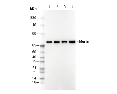 Lane 1: PC-3
Lane 2: MCF-7
Lane 3: Jrukat
Lane 4: SH-SY5Y