|
How to Cite 1. For In-Text Citation (Materials & Methods): 2. For Key Resources Table: |
||
|
Toll Free: (877) 796-6397 -- USA and Canada only -- |
Fax: +1-832-582-8590 Orders: +1-832-582-8158 |
Tech Support: +1-832-582-8158 Ext:3 Please provide your Order Number in the email. We strive to reply to |
Biological Description
| Specificity | Lck Antibody [P2J8] detects endogenous levels of total Lck protein. |
|---|---|
| Background | Lck is a Src family non‑receptor tyrosine kinase that localizes mainly to the inner leaflet of the plasma membrane of T lineage cells where it functions as a primary initiator and amplifier of T cell receptor signaling and contributes to additional receptor pathways that regulate development, activation, and effector function. The protein contains an N‑terminal myristoylation and palmitoylation region that targets it to membranes, followed by SH3 and SH2 domains that engage proline‑rich and phosphotyrosine‑containing motifs in partner proteins, and a C‑terminal kinase domain whose activity is controlled by intramolecular interactions governed by phosphorylation of the activation loop tyrosine and the C‑terminal inhibitory tyrosine. Association of Lck with the cytoplasmic tails of CD4 and CD8 co‑receptors positions the kinase adjacent to the TCR–CD3 complex during antigen recognition, where it phosphorylates immunoreceptor tyrosine‑based activation motifs within CD3 and ζ‑chains, creating docking sites for the Syk family kinase ZAP‑70 and initiating a cascade that recruits adaptor proteins such as LAT and SLP‑76 to assemble multi‑component signalosomes at the immune synapse. Catalytic activity of Lck integrates positive input from dephosphorylation of the C‑terminal inhibitory tyrosine and phosphorylation of the activation loop with negative regulation through Csk‑mediated phosphorylation and inhibitory protein interactions, establishing a tunable threshold for TCR signaling that influences thymic selection, peripheral activation, and sensitivity to antigen. Downstream of the TCR, Lck‑dependent phosphorylation events control multiple effector branches, including PLCγ1 activation with consequent calcium mobilization, Ras–MAPK pathway engagement, and activation of transcription factors such as NF‑AT, AP‑1, and NF‑κB, linking early membrane‑proximal kinase activity to gene expression programs that drive proliferation, differentiation, and cytokine production. The SH3 domain of Lck contributes specifically to coupling of proximal TCR phosphorylation to activation of the Ras–ERK arm of the MAPK pathway, illustrating that discrete structural modules within the kinase differentially regulate signaling outputs while leaving other branches such as calcium flux relatively intact when perturbed. Lck not only transduces TCR signals but also participates in co‑stimulatory and cytokine receptor pathways, including IL‑2 receptor signaling and CD2‑dependent activation, expanding its influence across multiple receptor systems that shape T cell expansion and functional polarization. Aberrant Lck activity or expression is linked to immunodeficiency, where insufficient signaling impairs thymocyte maturation and peripheral T cell responsiveness, and to autoimmunity and T cell malignancies, where excessive or deregulated activity contributes to pathological activation, chronic NF‑κB signaling, and oncogenic transformation. |
Usage Information
| Application | WB, IP, IHC, IF, FCM | Dilution |
|
||||||||||
|---|---|---|---|---|---|---|---|---|---|---|---|---|---|
| Reactivity | Mouse, Rat, Human | ||||||||||||
| Source | Rabbit Monoclonal Antibody | MW | 58 kDa | ||||||||||
| Storage Buffer | PBS, pH 7.2+50% Glycerol+0.05% BSA+0.01% NaN3 | Storage (from the date of receipt) |
-20°C (avoid freeze-thaw cycles), 2 years | ||||||||||
References
|
Application Data
WB
Validated by Selleck
-
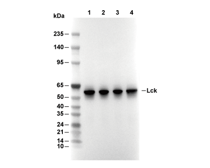 Lane 1: Raji, Lane 2: Ramos, Lane 3: Jurka, Lane 4: Mouse thymus