|
How to Cite 1. For In-Text Citation (Materials & Methods): 2. For Key Resources Table: |
||
|
Toll Free: (877) 796-6397 -- USA and Canada only -- |
Fax: +1-832-582-8590 Orders: +1-832-582-8158 |
Tech Support: +1-832-582-8158 Ext:3 Please provide your Order Number in the email. We strive to reply to |
Biological Description
| Specificity | Keratin Epithelial Antibody [J4M17] detects endogenous levels of total Keratin Epithelial protein. |
|---|---|
| Background | Keratin Epithelial proteins comprise type I (acidic) and type II (basic) intermediate filament components expressed across stratified and simple epithelia, forming obligate heterodimers that assemble into 10 nm filaments, providing mechanical resilience against shear stress and osmotic challenge. keratins organize central rod domains with 1A, 1B, 2A, and 2B alpha-helical segments staggered for protofilament polymerization alongside non-alpha-helical N- and C-terminal head/tail domains rich in glycine loops and arginine clusters that mediate lateral filament packing and desmin-plakoglobin interactions at desmosomes. Hyperproliferative signaling through EGFR-Ras-ERK cascades transcriptionally induces K6/K16/K17 expression via AP-1 and C/EBP factors that recruit 14-3-3σ to activate mTORC1 through Raptor phosphorylation, boosting protein synthesis and cell migration while K17 recruits 14-3-3ζ to plasma membrane Src-FAK complexes amplifying PI3K-AKT signaling for survival under wounding or inflammation. Barrier disruption triggers K6/K16/K17 upregulation through PKC-α mediated JNK activation that phosphorylates K17 serines, enhancing actin-myosin contractility and lamellipodia protrusion during reepithelialization, while K1/K10 pairs stabilize cornified envelopes through transglutaminase crosslinking with involucrin and loricrin. Absence of keratins impairs GLUT1/4 localization disrupting AMPK-mTOR balance and glucose uptake essential for epithelial energy homeostasis, with K14/K5 mutations causing epidermolysis bullosa simplex through filament aggregation and basal cell lysis. Epithelial keratins govern epidermal stratification, corneal wound healing, and glandular morphogenesis with differentiation-specific pairwise expression ensuring tissue integrity gradients. Dysregulation manifests in pachyonychia congenita and steatocystoma multiplex through K6a/K16/K17 mutations disrupting tonofilament anchorage. |
Usage Information
| Application | WB, IHC | Dilution |
|
||
|---|---|---|---|---|---|
| Reactivity | Rat, Chicken, Rabbit, Mouse, Human, Monkey, Bovine | ||||
| Source | Mouse Monoclonal Antibody | MW | 65-67 kDa, 52-59 kDa | ||
| Storage Buffer | PBS, pH 7.2+50% Glycerol+0.05% BSA+0.01% NaN3 | Storage (from the date of receipt) |
-20°C (avoid freeze-thaw cycles), 2 years | ||
References
|
Application Data
WB
Validated by Selleck
-
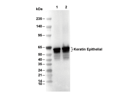 Lane 1: MCF7, Lane 2: A431