|
How to Cite 1. For In-Text Citation (Materials & Methods): 2. For Key Resources Table: |
||
|
Toll Free: (877) 796-6397 -- USA and Canada only -- |
Fax: +1-832-582-8590 Orders: +1-832-582-8158 |
Tech Support: +1-832-582-8158 Ext:3 Please provide your Order Number in the email. We strive to reply to |
Biological Description
| Specificity | IRF-7 Antibody [C15N21] detects endogenous levels of total IRF-7 protein. |
|---|---|
| Background | Interferon regulatory factor 7 (IRF-7) is a member of the IRF transcription factor family that binds specific DNA elements in interferon-stimulated promoters and integrates upstream innate immune receptor signaling into type I and III interferon gene expression, especially in plasmacytoid dendritic cells and lymphoid lineages, where its expression is enriched and inducible by virus, lipopolysaccharide, and type I interferons. The N-terminal region contains a conserved helix–turn–helix DNA-binding domain that recognizes interferon regulatory elements, while the C-terminal regulatory region carries multiple serine-rich motifs and interaction surfaces that control dimerization, nuclear import, and cofactor recruitment. Upstream activation begins at pattern-recognition receptors such as endosomal Toll-like receptors and cytosolic nucleic acid sensors, which signal through adaptor proteins including MyD88 or TRIF, TRAF family ubiquitin ligases, and TANK-binding kinase 1 or IKK-related kinases that phosphorylate IRF-7 on clustered serine residues in the C terminus. Phosphorylated IRF-7 forms homodimers or heterodimers with other IRFs, undergoes conformational exposure of its nuclear localization signal, accumulates in the nucleus, and assembles on interferon promoters together with chromatin-modifying coactivators to drive a broad antiviral transcriptional program. This transcriptional output reinforces interferon signaling through a positive feedback loop, shapes the amplitude and duration of type I interferon waves, and coordinates expression of numerous interferon-stimulated genes that regulate viral replication, antigen presentation, and inflammatory mediator production. Multiple post-translational modifications fine-tune this activity: phosphorylation marks correlate with activation, K63-linked ubiquitination promotes full transactivation, and other ubiquitin linkages or proteasomal targeting limit IRF-7 abundance and prevent excessive interferon output. IRF-7 also interfaces with NF-κB and AP-1 pathways at shared target promoters and participates in Epstein–Barr virus latency programs, where its induction and activation by latent membrane protein 1 link viral oncogenic signaling to interferon-related transcriptional networks. In physiological immunity, IRF-7 is critical for early systemic antiviral defense, shaping plasmacytoid dendritic cell function, influencing B cell responses, and contributing to broader orchestration of innate and adaptive immune communication. Dysregulated IRF-7 expression or activity alters interferon production thresholds, associates with autoimmune phenotypes characterized by chronic type I interferon signatures, and influences susceptibility or progression in infection, cancer, and other inflammation-associated pathologies where interferon balance is a major determinant of tissue outcome. |
Usage Information
| Application | WB, IP | Dilution |
|
||||
|---|---|---|---|---|---|---|---|
| Reactivity | Mouse, Rat, Hamster | ||||||
| Source | Rabbit Monoclonal Antibody | MW | 54 kDa | ||||
| Storage Buffer | PBS, pH 7.2+50% Glycerol+0.05% BSA+0.01% NaN3 | Storage (from the date of receipt) |
-20°C (avoid freeze-thaw cycles), 2 years | ||||
References
|
Application Data
WB
Validated by Selleck
-
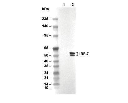 Lane 1: A20, Lane 2: A20 (mIFN-α, 10 ng/ml, 24 h)