|
How to Cite 1. For In-Text Citation (Materials & Methods): 2. For Key Resources Table: |
||
|
Toll Free: (877) 796-6397 -- USA and Canada only -- |
Fax: +1-832-582-8590 Orders: +1-832-582-8158 |
Tech Support: +1-832-582-8158 Ext:3 Please provide your Order Number in the email. We strive to reply to |
Biological Description
| Specificity | HOXA5 Antibody [C16C3] detects endogenous levels of total HOXA5 protein. |
|---|---|
| Background | HOXA5 belongs to the HOX family of homeobox transcription factors that establish positional identity along the anterior-posterior axis during embryonic development. This protein features a conserved 60-residue homeodomain with three alpha-helices that bind specific DNA sequences, flanked by N-terminal transactivation domains and a C-terminal hexapeptide motif for cofactor interactions. HOXA5 expression patterns the cervical-thoracic transition, appearing dynamically in somites, lateral plate mesoderm, lungs, kidneys, spinal cord, and developing musculoskeletal tissues. During organogenesis, HOXA5 directs skeletal patterning by regulating sclerotome differentiation into chondrocytes, osteoblasts, and perichondrium, while also shaping ribs, vertebrae, sternum, and forelimb girdle. It promotes alveolar myofibroblast specification in lungs through colocalization with PDGFRα, driving elastin deposition and septation essential for lung maturation. HOXA5-expressing cells contribute to tendons, ligaments, and dermis, influencing adhesion and morphogenesis of precartilaginous condensations. HOXA5 activates genes like Pax1 for acromion development and represses others to fine-tune vertebral identity and diaphragm formation. HOXA5 modulates Wnt/β-catenin by restraining WNT2B and NFATC1, promoting adipocyte differentiation via FABP4 upregulation and PKA/HSL inhibition. It intersects TGF-β pathways during tissue remodeling, enhancing anti-inflammatory signals and suppressing macrophage infiltration in adipose tissue. HOXA5 loss correlates with lung tumorigenesis by impairing p53-mediated apoptosis and proliferation control, while restored expression curbs metastasis. Hoxa5-null mice exhibit respiratory failure from defective tracheoesophageal septation, craniofacial anomalies, and female reproductive tract malformations. Aberrant silencing associates with hypertrophic obesity, insulin resistance, and chronic inflammation through dysregulated adipogenesis. |
Usage Information
| Application | WB | Dilution |
|
||
|---|---|---|---|---|---|
| Reactivity | Human | ||||
| Source | Rabbit Monoclonal Antibody | MW | 29 kDa | ||
| Storage Buffer | PBS, pH 7.2+50% Glycerol+0.05% BSA+0.01% NaN3 | Storage (from the date of receipt) |
-20°C (avoid freeze-thaw cycles), 2 years | ||
| WB |
Experimental Protocol:
Sample preparation
1. Tissue: Lyse the tissue sample by adding an appropriate volume of ice-cold RIPA/Nuclear Lysis Buffer (containing Protease Inhibitor Cocktail),and homogenize the tissue at a low temperature. 2. Adherent cell: Aspirate the culture medium and wash the cells with ice-cold PBS twice. Lyse the cells by adding an appropriate volume of RIPA/Nuclear Lysis Buffer (containing Protease Inhibitor Cocktail) and put the sample on ice for 5 min. 3. Suspension cell: Transfer the culture medium to a pre-cooled centrifuge tube. Centrifuge and aspirate the supernatant. Wash the cells with ice-cold PBS twice. Lyse the cells by adding an appropriate volume of RIPA/Nuclear Lysis Buffer (containing Protease Inhibitor Cocktail) and put the sample on ice for 5 min. 4. Place the lysate into a pre-cooled microcentrifuge tube. Centrifuge at 4°C for 15 min. Collect the supernatant;
5. Remove a small volume of lysate to determine the protein concentration;
6. Combine the lysate with protein loading buffer. Boil 20 µL sample under 95-100°C for 5 min. Centrifuge for 5 min after cool down on ice.
2. Power up 80V for 30 minutes. Then the power supply is adjusted (110 V~150 V), the Marker is observed, and the electrophoresis can be stopped when the indicator band of the predyed protein Marker where the protein is located is properly separated. (Note that the current should not be too large when electrophoresis, too large current (more than 150 mA) will cause the temperature to rise, affecting the result of running glue. If high currents cannot be avoided, an ice bath can be used to cool the bath.)
Transfer membrane
1. Take out the converter, soak the clip and consumables in the pre-cooled converter;
2. Activate PVDF membrane with methanol for 1 min and rinse with transfer buffer;
3. Install it in the order of "black edge of clip - sponge - filter paper - filter paper - glue -PVDF membrane - filter paper - filter paper - sponge - white edge of clip"; 4. The protein was electrotransferred to PVDF membrane. ( 0.45 µm PVDF membrane is recommended ) Reference Table for Selecting PVDF Membrane Pore Size Specifications Recommended conditions for wet transfer: 200 mA, 60 min. ( Note that the transfer conditions can be adjusted according to the protein size. For high-molecular-weight proteins, a higher current and longer transfer time are recommended. However, ensure that the transfer tank remains at a low temperature to prevent gel melting.)
Block
1. After electrotransfer, wash the film with TBST at room temperature for 5 minutes;
2. Incubate the film in the blocking solution for 1 hour at room temperature;
3. Wash the film with TBST for 3 times, 5 minutes each time.
Antibody incubation
1. Use 5% skim milk powder to prepare the primary antibody working liquid (recommended dilution ratio for primary antibody 1:1000), gently shake and incubate with the film at 4°C overnight; 2. Wash the film with TBST 3 times, 5 minutes each time;
3. Add the secondary antibody to the blocking solution and incubate with the film gently at room temperature for 1 hour;
4. After incubation, wash the film with TBST 3 times for 5 minutes each time.
Antibody staining
1. Add the prepared ECL luminescent substrate (or select other color developing substrate according to the second antibody) and mix evenly;
2. Incubate with the film for 1 minute, remove excess substrate (keep the film moist), wrap with plastic film, and expose in the imaging system.
|
References
|
Application Data
WB
Validated by Selleck
-
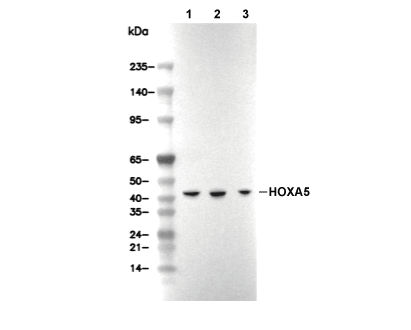 Lane 1: 293T, Lane 2: SH-SY5Y, Lane 3: Caco-2