|
How to Cite 1. For In-Text Citation (Materials & Methods): 2. For Key Resources Table: |
||
|
Toll Free: (877) 796-6397 -- USA and Canada only -- |
Fax: +1-832-582-8590 Orders: +1-832-582-8158 |
Tech Support: +1-832-582-8158 Ext:3 Please provide your Order Number in the email. We strive to reply to |
Biological Description
| Specificity | HLA-DR Antibody [L16M22] detects endogenous levels of total HLA-DR protein. |
|---|---|
| Background | HLA‑DR is a heterodimeric class II major histocompatibility complex receptor composed of an invariant DRα chain and a polymorphic DRβ chain that localizes to the surface of professional antigen‑presenting cells and mediates presentation of exogenous and selected endogenous peptides to CD4 T cells, placing it at the core of adaptive immune initiation and regulation. The extracellular portion of each chain contains two Ig‑like domains that pair to form an open‑ended peptide‑binding groove supported by a β‑sheet floor and flanking α‑helices, with key peptide contact residues concentrated in the N‑terminal portions of both chains and specificity largely dictated by polymorphic positions in DRβ. Peptides of roughly 10–30 residues generated from endocytosed proteins in late endosomes and lysosomes are loaded onto newly synthesized HLA‑DR in specialized MHC class II compartments; an invariant chain initially occupies the groove and targets the complex to the endocytic pathway, where sequential proteolysis leaves CLIP fragments that are exchanged for higher‑affinity peptides under the control of the DM chaperone. The resulting HLA‑DR–peptide complexes traffic to the plasma membrane and engage TCRs on CD4 T cells, providing signal 1 for T‑cell activation, while co‑stimulatory receptors and cytokines supplied by the antigen‑presenting cell determine the differentiation and effector profile of the responding T‑cell population. In peripheral tissues and tumor microenvironments, HLA‑DR presents peptides derived from internalized microbial antigens, self‑proteins, and tumor‑associated proteins taken up by phagocytosis or macropinocytosis, guiding helper T‑cell responses that support antibody production, macrophage activation, and cytotoxic T‑cell priming directed against pathogens or transformed cells. Autophagy‑linked processing routes deliver cytosolic and nuclear proteins to autolysosomes for loading onto HLA‑DR, a mechanism that contributes to positive and negative selection of CD4 T cells in the thymus and to maintenance of central tolerance by presenting a broader self‑peptide repertoire. Expression of HLA‑DR is largely restricted to dendritic cells, macrophages, B cells, and thymic epithelial cells under homeostatic conditions but is inducible on additional cell types under inflammatory stimulation, making surface HLA‑DR a marker of immune activation and antigen‑presenting cell maturation. Extensive polymorphism in HLA‑DRB1 and linked DRB loci shapes the peptide‑binding groove and TCR contact surfaces, creating distinct peptide‑binding motifs that influence susceptibility or resistance to autoimmune diseases, infection outcomes, and responses to vaccines and biologic therapies. HLA‑DR matching between donor and recipient is a major determinant of graft acceptance and long‑term survival in hematopoietic and solid‑organ transplantation, reflecting the strong alloreactive T‑cell responses elicited by mismatched DR–peptide complexes. |
Usage Information
| Application | WB, IHC, IF | Dilution |
|
||||||
|---|---|---|---|---|---|---|---|---|---|
| Reactivity | Human | ||||||||
| Source | Rabbit Monoclonal Antibody | MW | 29 kDa | ||||||
| Storage Buffer | PBS, pH 7.2+50% Glycerol+0.05% BSA+0.01% NaN3 | Storage (from the date of receipt) |
-20°C (avoid freeze-thaw cycles), 2 years | ||||||
References
|
Application Data
WB
Validated by Selleck
-
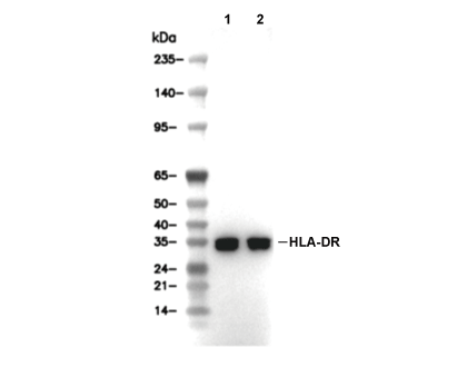 Lane 1: Raji, Lane 2: Ramos