|
Toll Free: (877) 796-6397 -- USA and Canada only -- |
Fax: +1-832-582-8590 Orders: +1-832-582-8158 |
Tech Support: +1-832-582-8158 Ext:3 Please provide your Order Number in the email. |
Technical Data
| Formula | C15H22N2O2 |
||||||
| Molecular Weight | 262.35 | CAS No. | 347174-05-4 | ||||
| Solubility (25°C)* | In vitro | DMSO | 52 mg/mL (198.2 mM) | ||||
| Ethanol | 52 mg/mL (198.2 mM) | ||||||
| Water | Insoluble | ||||||
| In vivo (Add solvents to the product individually and in order) |
|
||||||
|
* <1 mg/ml means slightly soluble or insoluble. * Please note that Selleck tests the solubility of all compounds in-house, and the actual solubility may differ slightly from published values. This is normal and is due to slight batch-to-batch variations. * Room temperature shipping (Stability testing shows this product can be shipped without any cooling measures.) |
|||||||
Preparing Stock Solutions
Biological Activity
| Description | Fer-1 (Ferrostatin-1) is a potent and selective inhibitor of ferroptosis with EC50 of 60 nM. | ||
|---|---|---|---|
| Targets |
|
||
| In vitro | Ferrostatin-1 (2 μM) prevents erastin-induced ferroptosis in cancer cells, as well as glutamate-induced cell death in postnatal rat brain slices. Ferrostatin-1 is a lipid ROS scavenger, with the N-cyclohexyl moiety serving as a lipo-philic anchor within biological membranes. Ferrostatin-1 does not inhibit extracellular signal -regulated kinase (ERK) phos-phorylation or arrest the proliferation of HT-1080 cells, suggesting that it does not inhibit the MEK/ERK pathway, chelate iron, or inhibit protein synthesis. Ferrostatin-1 does, however, prevent erastin-induced accumulation of cytosolic and lipid ROS. Ferrostatin-1 readily oxidizes the stable radical 2,2-diphenyl-1-picrylhydrazyl (DPPH) under cell-free conditions. [1] |
||
| In vivo | Ferrostatin-1 (Fer-1), a synthetic antioxidant, is a potent and selective inhibitor of ferroptosis. It acts via a reductive mechanism to prevent damage to membrane lipids and thereby inhibits cell death.[2] |
Protocol (from reference)
| Cell Assay: |
|
|---|---|
| Animal Study: |
|
References
|
Customer Product Validation
-
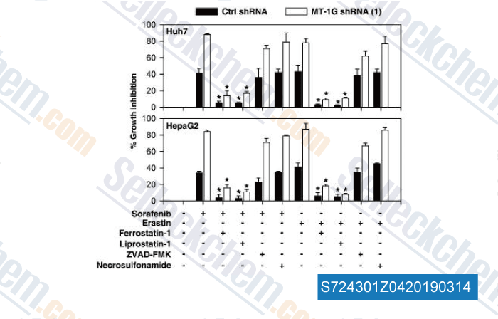
Data from [ , , Hepatology, 2016, 64(2):488-500 ]
-
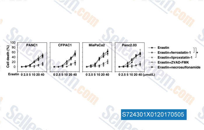
Data from [ , , Cancer Res, 2017, 77(8):2064-2077 ]
-
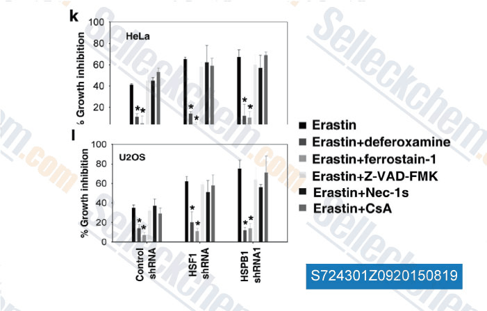
Data from [ , , Oncogene, 2015, 10.1038/onc.2015.32 ]
-
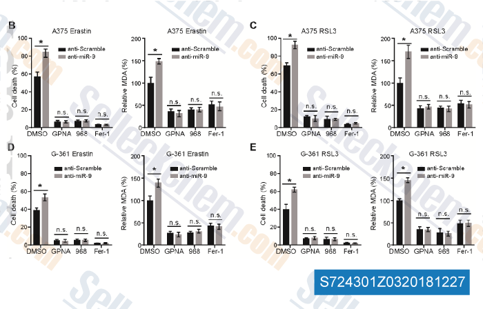
Data from [ , , Mol Carcinog, 2018, 57(11):1566-1576 ]
Selleck's Fer-1 (Ferrostatin-1) has been cited by 667 publications
| Lymphoma accelerates T cell and tissue aging [ Cancer Cell, 2025, S1535-6108(25)00329-0] | PubMed: 40845845 |
| Radiotherapy promotes cuproptosis and synergizes with cuproptosis inducers to overcome tumor radioresistance [ Cancer Cell, 2025, S1535-6108(25)00132-1] | PubMed: 40215978 |
| Cytosolic cytochrome c represses ferroptosis [ Cell Metab, 2025, S1550-4131(25)00149-4] | PubMed: 40233758 |
| RIPK1 senses S-adenosylmethionine scarcity to drive cell death and inflammation [ Cell Metab, 2025, S1550-4131(25)00294-3] | PubMed: 40570842 |
| Harnessing the FGFR2/NF2/YAP signaling-dependent necroptosis to develop an FGFR2/IL-8 dual blockade therapeutic strategy [ Nat Commun, 2025, 16(1):4128] | PubMed: 40319089 |
| Senescence-associated lysosomal dysfunction impairs cystine deprivation-induced lipid peroxidation and ferroptosis [ Nat Commun, 2025, 16(1):6617] | PubMed: 40731111 |
| Obesity-associated macrophages dictate adipose stem cell ferroptosis and visceral fat dysfunction by propagating mitochondrial fragmentation [ Nat Commun, 2025, 16(1):7564] | PubMed: 40813577 |
| S100P is a ferroptosis suppressor to facilitate hepatocellular carcinoma development by rewiring lipid metabolism [ Nat Commun, 2025, 16(1):509] | PubMed: 39779666 |
| Iron supplementation alleviates pathologies in a mouse model of facioscapulohumeral muscular dystrophy [ J Clin Invest, 2025, e181881] | PubMed: 40591405 |
| PIP5K1A Suppresses Ferroptosis and Induces Sorafenib Resistance by Stabilizing NRF2 in Hepatocellular Carcinoma [ Adv Sci (Weinh), 2025, 12(30):e04372] | PubMed: 40405713 |
RETURN POLICY
Selleck Chemical’s Unconditional Return Policy ensures a smooth online shopping experience for our customers. If you are in any way unsatisfied with your purchase, you may return any item(s) within 7 days of receiving it. In the event of product quality issues, either protocol related or product related problems, you may return any item(s) within 365 days from the original purchase date. Please follow the instructions below when returning products.
SHIPPING AND STORAGE
Selleck products are transported at room temperature. If you receive the product at room temperature, please rest assured, the Selleck Quality Inspection Department has conducted experiments to verify that the normal temperature placement of one month will not affect the biological activity of powder products. After collecting, please store the product according to the requirements described in the datasheet. Most Selleck products are stable under the recommended conditions.
NOT FOR HUMAN, VETERINARY DIAGNOSTIC OR THERAPEUTIC USE.