|
How to Cite 1. For In-Text Citation (Materials & Methods): 2. For Key Resources Table: |
||
|
Toll Free: (877) 796-6397 -- USA and Canada only -- |
Fax: +1-832-582-8590 Orders: +1-832-582-8158 |
Tech Support: +1-832-582-8158 Ext:3 Please provide your Order Number in the email. We strive to reply to |
Biological Description
| Specificity | Fascin Antibody [K2B22] detects endogenous levels of total Fascin protein. |
|---|---|
| Background | Fascin stands as the principal actin-bundling protein within the filopodial actin-crosslinking family, essential for organizing parallel actin filaments into compact bundles that drive the extension of cellular protrusions such as filopodia and lamellipodia. Its monomeric globular architecture incorporates four β-trefoil folds, with two conserved actin-binding sites, one in the N-terminal β-trefoil domain 1 featuring Ser39 susceptible to protein kinase C phosphorylation, and another in β-trefoil domain 3, that enable high-affinity, charge-neutralizing interactions with F-actin, promoting lateral filament association at a stoichiometry of one fascin per actin dimer while maintaining bundle flexibility for dynamic protrusion growth. Phosphorylation at Ser39 disrupts the first binding site, reducing bundling efficiency and filopodia formation, whereas dephosphorylated fascin stabilizes bundles to facilitate force generation during migration. Fascin engages the armadillo-repeat domain of β-catenin both in vitro and in vivo, colocalizing with β-catenin and cadherins at leading edges to reinforce adhesion turnover, and activates Wnt/β-catenin signaling via focal adhesion kinase (FAK) dependence, where FAK inhibition blocks fascin-induced nuclear β-catenin accumulation and downstream target expression like c-Myc and cyclin D1. This pathway sustains epithelial-mesenchymal transition by elevating vimentin and N-cadherin while suppressing E-cadherin, enhancing collective cell migration and stem cell-like properties in tumorspheres. Expressed at low levels in normal epithelia, fascin localizes to cytoplasmic bundles in migratory neurons, dendritic cells, and vascular smooth muscle, but upregulation occurs through Sp1 promoter binding enhanced by EGFR-MAPK/ERK signaling. Overexpression correlates with lymph node metastasis, advanced staging, and poor prognosis across breast, colorectal, esophageal squamous cell, prostate, and ovarian carcinomas, where it drives invasion independently of its bundling role in some contexts. |
Usage Information
| Application | WB, IP | Dilution |
|
||||
|---|---|---|---|---|---|---|---|
| Reactivity | Human, Mouse, Rat, Monkey | ||||||
| Source | Mouse Monoclonal Antibody | MW | 54 kDa | ||||
| Storage Buffer | PBS, pH 7.2+50% Glycerol+0.05% BSA+0.01% NaN3 | Storage (from the date of receipt) |
-20°C (avoid freeze-thaw cycles), 2 years | ||||
References
|
Application Data
WB
Validated by Selleck
-
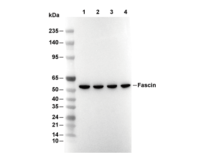 Lane 1: ACHN, Lane 2: Hela, Lane 3: HepG2, Lane 4: BT-549