|
How to Cite 1. For In-Text Citation (Materials & Methods): 2. For Key Resources Table: |
||
|
Toll Free: (877) 796-6397 -- USA and Canada only -- |
Fax: +1-832-582-8590 Orders: +1-832-582-8158 |
Tech Support: +1-832-582-8158 Ext:3 Please provide your Order Number in the email. We strive to reply to |
Biological Description
| Specificity | Fas Antibody [D2F4] detects endogenous levels of total Fas protein. |
|---|---|
| Background | Fas (CD95/Apo-1), a prototypical death receptor of the TNF receptor superfamily, transduces extrinsic apoptosis signals critical for immune homeostasis, lymphocyte deletion, and tumor surveillance through trimerization induced by multimeric FasL binding to its extracellular cysteine-rich domains. Ligand engagement recruits the adaptor FADD via death domain interactions, forming the death-inducing signaling complex (DISC) that docks procaspase-8 through DED-mediated oligomerization, enabling autocatalytic activation independent of mitochondrial amplification in type I cells or BID cleavage in type II cells to converge on effector caspase-3/7 execution. Membrane-bound FasL from activated CD8 T cells and NK cells potently clusters Fas trimers for full DISC assembly, whereas soluble FasL requires secondary aggregation for physiological potency, distinguishing it from agonistic antibodies that variably signal death versus survival through NF-κB/ERK non-apoptotic branches. This pathway operates stringently downstream of FADD/caspase-8 as evidenced by complete blockade in deficient cells, while remaining insensitive to Bcl-2/Bcl-xL mitochondrial regulation, establishing Fas as the dominant extrinsic apoptosis trigger in lymphocytes and hepatocytes. Physiologically, Fas governs activation-induced cell death to prevent autoimmunity, contracts immune responses post-infection, and prunes double-negative thymocytes, making it indispensable for researchers quantifying effector deletion via Annexin V or dissecting tolerance breakdown in lpr/gld models. Tumors exploit FasL expression as a counterattack mechanism, killing Fas-sensitive TILs while acquiring caspase-8 mutations for resistance, though FasL suppression paradoxically enhances tumor clearance through restored lymphocyte infiltration. Immune-privileged sites weaponize constitutive FasL to exclude Fas-bearing leukocytes. |
Usage Information
| Application | WB, IHC, IF | Dilution |
|
||||||
|---|---|---|---|---|---|---|---|---|---|
| Reactivity | Human | ||||||||
| Source | Rabbit Monoclonal Antibody | MW | 37 kDa | ||||||
| Storage Buffer | PBS, pH 7.2+50% Glycerol+0.05% BSA+0.01% NaN3 | Storage (from the date of receipt) |
-20°C (avoid freeze-thaw cycles), 2 years | ||||||
References
|
Application Data
WB
Validated by Selleck
-
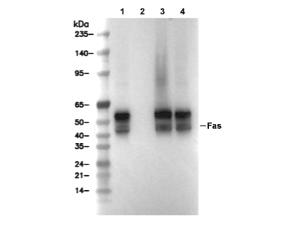 Lane 1: Hela, Lane 2: Hela (KO Fas), Lane 3: Raji, Lane 4: Ramos