|
How to Cite 1. For In-Text Citation (Materials & Methods): 2. For Key Resources Table: |
||
|
Toll Free: (877) 796-6397 -- USA and Canada only -- |
Fax: +1-832-582-8590 Orders: +1-832-582-8158 |
Tech Support: +1-832-582-8158 Ext:3 Please provide your Order Number in the email. We strive to reply to |
Technical Data
| Formula | C22H23N3O4 |
||||||
| Molecular Weight | 393.44 | CAS No. | 183321-74-6 | ||||
| Solubility (25°C)* | In vitro | DMSO | 100 mg/mL (254.16 mM) | ||||
| Ethanol | 15 mg/mL (38.12 mM) | ||||||
| Water | Insoluble | ||||||
| In vivo (Add solvents to the product individually and in order) |
|
||||||
|
* <1 mg/ml means slightly soluble or insoluble. * Please note that Selleck tests the solubility of all compounds in-house, and the actual solubility may differ slightly from published values. This is normal and is due to slight batch-to-batch variations. * Room temperature shipping (Stability testing shows this product can be shipped without any cooling measures.) |
|||||||
Preparing Stock Solutions
Biological Activity
| Description | Erlotinib is an EGFR inhibitor with IC50 of 2 nM, >1000-fold more sensitive for EGFR than human c-Src or v-Abl. Erlotinib induces autophagy. | ||
|---|---|---|---|
| Targets |
|
||
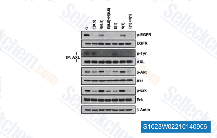
| In vitro | Erlotinib HCl potently inhibits EGFR activation in intact cells including HNS human head and neck tumor cells (IC50 20nM), DiFi human colon cancer cells and MDA MB-468 human breast cancer cells. This compound (1 μM) induces apoptosis in DiFi human colon cancer cells. It inhibits growth of a panel of NSCLC cell lines including A549, H322, H3255, H358 H661, H1650, H1975, H1299, H596 with IC50 ranging from 29 nM to >20 μM. This chemical (2 μM) significantly inhibits growth of AsPC-1 and BxPC-3 pancreatic cells. The effects of this compound in combination with gemcitabine are considered additive in KRAS-mutated pancreatic cancer cells. Ten micromolar of this chemical inhibits EGFR phospho-rylation at the Y845 (Src-dependent phosphorylation) and Y1068 (auto-phosphorylation) sites. Combination with this compound could down-modulate rapamycin-stimulated Akt activity and produces a synergistic effect on cell growth inhibition. |
||
| In vivo | At doses of 100 mg/kg, Erlotinib HCl completely prevents EGF-induced autophosphorylation of EGFR in human HN5 tumors growing as xenografts in athymic mice and of the hepatic EGFR of the treated mice. This compound (100 mg/Kg) inhibits H460a and A549 tumor models with 71 and 93% inhibition rate. |
Protocol (from reference)
| Kinase Assay: |
|
|---|---|
| Cell Assay: |
|
| Animal Study: |
|
References
|
Customer Product Validation
-
Data from [ Cancer Res , 2014 , 4(1):253-62 ]
-
Data from [ Head Neck , 2013 , 35, 86-93 ]
-
Data from [ Tuberc Respir Dis , 2013 , 75(1), 9-17 ]
-
Data from [ Nat Genet , 2012 , 44(8):852-60 ]
Selleck's Erlotinib (CP-358774) Has Been Cited by 653 Publications
| Non-canonical dihydrolipoyl transacetylase promotes chemotherapy resistance via mitochondrial tetrahydrofolate signaling [ Nat Commun, 2025, 16(1):8932] | PubMed: 41062483 |
| EGFR TKIs suppress MUC1 glycosylation through the PI3K/AKT/SP1/C1GALT1 pathway to enhance TnMUC1 CAR-T efficacy in EGFR-mutant NSCLC [ Cell Rep Med, 2025, S2666-3791(25)00272-1] | PubMed: 40562040 |
| Multi-layer stratified oncology platform utilizing transcriptomics, prostate cancer organoids, and modeling of drug response [ J Exp Clin Cancer Res, 2025, 44(1):290] | PubMed: 41094672 |
| AP1-mediated reprogramming of EGFR expression triggers resistance to BLU-667 and LOXO-292 in RET-rearranged tumors [ J Exp Clin Cancer Res, 2025, 44(1):154] | PubMed: 40405293 |
| Targeting CDK12/13 Drives Mitotic Arrest to Overcome Resistance to KRASG12C Inhibitors [ Cancer Res, 2025, 10.1158/0008-5472.CAN-25-0450] | PubMed: 41165466 |
| Anti-galectin-9 therapy synergizes with EGFR inhibition to reprogram the tumor microenvironment and overcome immune evasion [ J Immunother Cancer, 2025, 13(7)e010926] | PubMed: 40664443 |
| Oncogenic RARγ isoforms promote head and neck cancer proliferation through vinexin-β-mediated cell cycle acceleration and autocrine activation of EGFR signal [ Int J Biol Sci, 2025, 21(1):1-16] | PubMed: 39744424 |
| The AURKA inhibitor alters the immune microenvironment and enhances targeting B7-H3 immunotherapy in glioblastoma [ JCI Insight, 2025, e173700] | PubMed: 39928563 |
| Rare mutations at EGFR L747 position: molecular characteristics and superior response to afatinib in NSCLC patients [ ESMO Open, 2025, 10(9):105558] | PubMed: 40834500 |
| Targeting both wild-type EGFR and its drug-resistant mutants with erlotinib-aptamer conjugates [ Eur J Med Chem, 2025, 296:117871] | PubMed: 40554308 |
RETURN POLICY
Selleck Chemical’s Unconditional Return Policy ensures a smooth online shopping experience for our customers. If you are in any way unsatisfied with your purchase, you may return any item(s) within 7 days of receiving it. In the event of product quality issues, either protocol related or product related problems, you may return any item(s) within 365 days from the original purchase date. Please follow the instructions below when returning products.
SHIPPING AND STORAGE
Selleck products are transported at room temperature. If you receive the product at room temperature, please rest assured, the Selleck Quality Inspection Department has conducted experiments to verify that the normal temperature placement of one month will not affect the biological activity of powder products. After collecting, please store the product according to the requirements described in the datasheet. Most Selleck products are stable under the recommended conditions.
NOT FOR HUMAN, VETERINARY DIAGNOSTIC OR THERAPEUTIC USE.