|
Toll Free: (877) 796-6397 -- USA and Canada only -- |
Fax: +1-832-582-8590 Orders: +1-832-582-8158 |
Tech Support: +1-832-582-8158 Ext:3 Please provide your Order Number in the email. |
Biological Description
| Specificity | DKK1 Rabbit mAb recognizes total endogenous levels of DKK1 protein. |
|---|---|
| Background | The Dickkopf (DKK) family consists of glycoproteins ranging from 255 to 350 amino acids, with four members identified in vertebrates: DKK1, DKK2, DKK3, and DKK4. Each DKK protein contains two conserved cysteine-rich domains that facilitate protein-protein interactions, and their expression is tightly regulated both spatially and temporally during development. DKKs bind with high affinity to Kremen1 and Kremen2, transmembrane proteins that also regulate Wnt signaling. Among the DKK family, DKK1 is the most extensively studied. DKK1 is a 266-amino acid protein with a molecular weight of approximately 26 kDa, characterized by six secondary structures, including two alpha helices and four beta-sheets. DKK1 is preferentially expressed in certain adult tissues, such as bone, placenta, prostate, spleen, and colon, and it plays a crucial role as an antagonist of the Wnt signaling pathway. It is involved in embryonic development, inducing head formation and limb morphogenesis. By inhibiting Wnt signaling, DKK1 promotes heart muscle formation in the anterior lateral mesoderm while suppressing erythropoiesis. Elevated levels of DKK1 are observed in various cancers, including lung cancer, esophageal squamous cell carcinoma, and hormone-resistant breast cancer, whereas decreased expression is found in malignant melanoma and colorectal cancer. |
Usage Information
| Application | WB, IHC | Dilution |
|
||||
|---|---|---|---|---|---|---|---|
| Reactivity | Human | ||||||
| Source | Rabbit | MW | 29 kDa | ||||
| Storage Buffer | PBS, pH 7.2+50% Glycerol+0.05% BSA+0.01% NaN₃ | Storage (from the date of receipt) |
–20°C (avoid freeze-thaw cycles), 2 years | ||||
| WB |
Experimental Protocol:
Sample preparation
1. Tissue: Lyse the tissue sample by adding an appropriate volume of ice-cold RIPA/NP-40 Lysis Buffer (containing Protease Inhibitor Cocktail),and homogenize the tissue at a low temperature or lyse it by sonication on ice, then incubate on ice for 30 minutes. 2. Adherent cell: Aspirate the culture medium and transfer the cells into an EP tube. Wash the cells with ice-cold PBS twice. Add an appropriate volume of RIPA/NP-40 Lysis Buffer (containing Protease Inhibitor Cocktail), sonicate to lyse the cells, and incubate on ice for 30 minutes. 3. Suspension cell: Transfer the culture medium to a pre-cooled centrifuge tube. Centrifuge and aspirate the supernatant. Wash the cells with ice-cold PBS twice.Add an appropriate volume of RIPA/NP-40 Lysis Buffer (containing Protease Inhibitor Cocktail), sonicate to lyse the cells, and incubate on ice for 30 minutes. 4. Place the lysate into a pre-cooled microcentrifuge tube. Centrifuge at 4°C for 15 min. Collect the supernatant;
5. Remove a small volume of lysate to determine the protein concentration;
6. Combine the lysate with protein loading buffer. Boil 20 µL sample under 95-100°C for 5 min. Centrifuge for 5 min after cool down on ice.
Electrophoretic separation
1. According to the concentration of extracted protein, load appropriate amount of protein sample and marker onto SDS-PAGE gels for electrophoresis. Recommended separating gel (lower gel) concentration: 10%. Reference Table for Selecting SDS-PAGE Separation Gel Concentrations 2. Power up 80V for 30 minutes. Then the power supply is adjusted (110 V~150 V), the Marker is observed, and the electrophoresis can be stopped when the indicator band of the predyed protein Marker where the protein is located is properly separated. (Note that the current should not be too large when electrophoresis, too large current (more than 150 mA) will cause the temperature to rise, affecting the result of running glue. If high currents cannot be avoided, an ice bath can be used to cool the bath.)
Transfer membrane
1. Take out the converter, soak the clip and consumables in the pre-cooled converter;
2. Activate PVDF membrane with methanol for 1 min and rinse with transfer buffer;
3. Install it in the order of "black edge of clip - sponge - filter paper - filter paper - glue -PVDF membrane - filter paper - filter paper - sponge - white edge of clip"; 4. The protein was electrotransferred to PVDF membrane. ( 0.45 µm PVDF membrane is recommended ) Reference Table for Selecting PVDF Membrane Pore Size Specifications Recommended conditions for wet transfer: 200 mA, 60 min. ( Note that the transfer conditions can be adjusted according to the protein size. For high-molecular-weight proteins, a higher current and longer transfer time are recommended. However, ensure that the transfer tank remains at a low temperature to prevent gel melting.)
Block
1. After electrotransfer, wash the film with TBST at room temperature for 5 minutes;
2. Incubate the film in the blocking solution for 1 hour at room temperature;
3. Wash the film with TBST for 3 times, 5 minutes each time.
Antibody incubation
1. Use 5% skim milk powder to prepare the primary antibody working liquid (recommended dilution ratio for primary antibody 1:1000), gently shake and incubate with the film at 4°C overnight; 2. Wash the film with TBST 3 times, 5 minutes each time;
3. Add the secondary antibody to the blocking solution and incubate with the film gently at room temperature for 1 hour;
4. After incubation, wash the film with TBST 3 times for 5 minutes each time.
Antibody staining
920. Add the prepared ECL luminescent substrate (or select other color developing substrate according to the second antibody) and mix evenly;
2. Incubate with the film for 1 minute, remove excess substrate (keep the film moist), wrap with plastic film, and expose in the imaging system.
|
| WB |
Western Blotting
Sample preparation
1. Aspirate media from cultures and Wash the cells with 1X PBS.
2. Lyse cells by adding 1X SDS sample buffer and transfer the extract to a microcentrifuge tube. Keep onice.
3. Sonicate for 10–15 sec to complete cell lysis and shear DNA.
4. Heat a 20 µl sample to 95–100°C for 5 min, then cool on ice.
5. Centrifuge for 5 min (with Microcentrifuge).
6. Load appropriate volumes of samples onto SDS-PAGE gel (loading quantity of protein sample depends on the concentration of extracted proteins).
NOTE: At the same time, please load the pre-stained molecular weight markers to determine molecular weights and verify electrotransfer.
7. Electrotransfer to nitrocellulose/PVDF membrane (For wet transfer: 150mA 90min).
Membrane Blocking and Antibody Incubations
a. Blocking
1. (Optional) After transfer, wash the transferred membrane with TBS for 5 min at room temperature.
2. Incubate the membrane in the blocking buffer for 1 hr at room temperature.
3. Wash three times for 5 min each with TBST.
b. Antibodies Incubation
1. Incubate membrane and primary antibody (at the appropriate dilution and diluent recommended) in a primary antibody dilution buffer with gentle agitation overnight at 4°C.
2. Wash three times for 5 min each with TBST.
3. Incubate membrane with an appropriate second antibodydissolved in the blocking buffer with gentle agitation for 1 hr at room temperature.
4. Wash three times for 5 min each with TBST.
5. Proceed with detection.
Detection of Proteins
1. After antibodies incubation, Wash membrane three times for 5 minutes in TBST.
2. PrepareECL Reagent (or other chromogenic agents/substrate according to your second antibody). Mix well.
3. Incubate substrate with membrane for 1 minute, remove excess solution (membrane remains wet), wrap in plastic and expose in the imaging system.
|
| IHC |
Experimental Protocol:
Deparaffinization/Rehydration
1. Deparaffinize/hydrate sections:
2. Incubate sections in three washes of xylene for 5 min each.
3. Incubate sections in two washes of 100% ethanol for 10 min each.
4. Incubate sections in two washes of 95% ethanol for 10 min each.
5. Wash sections two times in dH2O for 5 min each.
6.Antigen retrieval: For Citrate: Heat slides in a microwave submersed in 1X citrate unmasking solution until boiling is initiated; continue with 10 min at a sub-boiling temperature (95°-98°C). Cool slides on bench top for 30 min.
Staining
1. Wash sections in dH2O three times for 5 min each.
2. Incubate sections in 3% hydrogen peroxide for 10 min.
3. Wash sections in dH2O two times for 5 min each.
4. Wash sections in wash buffer for 5 min.
5. Block each section with 100–400 µl of blocking solution for 1 hr at room temperature.
6. Remove blocking solution and add 100–400 µl primary antibody diluent in to each section. Incubate overnight at 4°C.
7. Remove antibody solution and wash sections with wash buffer three times for 5 min each.
8. Cover section with 1–3 drops HRPas needed. Incubate in a humidified chamber for 30 min at room temperature.
9. Wash sections three times with wash buffer for 5 min each.
10. Add DAB Chromogen Concentrate to DAB Diluent and mix well before use.
11. Apply 100–400 µl DAB to each section and monitor closely. 1–10 min generally provides an acceptable staining intensity.
12. Immerse slides in dH2O.
13. If desired, counterstain sections with hematoxylin.
14. Wash sections in dH2O two times for 5 min each.
15. Dehydrate sections: Incubate sections in 95% ethanol two times for 10 sec each; Repeat in 100% ethanol, incubating sections two times for 10 sec each; Repeat in xylene, incubating sections two times for 10 sec each.
16. Mount sections with coverslips and mounting medium.
|
Application Data
WB
Validated by Selleck
-
 Lane 1: A549
Lane 2: PC-3
Lane 3: DU145
Lane 4: SK-OV-3
IHC
Validated by Selleck
-
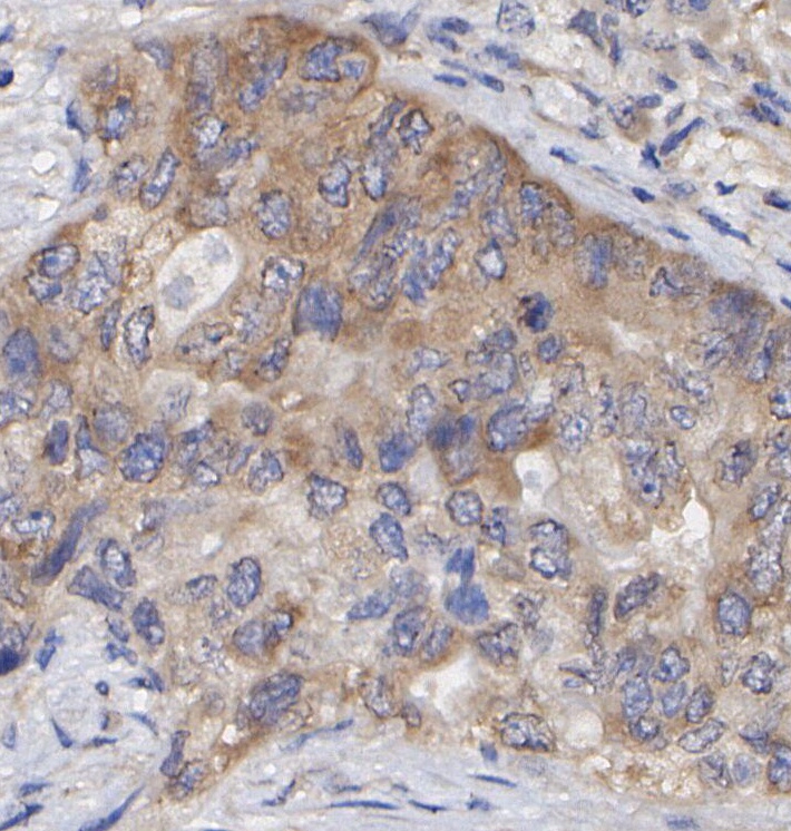 Immunohistochemical analysis of formalin fixed paraffin embedded human Lung adenocarcinoma tissue with F1661 at 1/100 dilution.