
|
How to Cite 1. For In-Text Citation (Materials & Methods): 2. For Key Resources Table: |
||
|
Toll Free: (877) 796-6397 -- USA and Canada only -- |
Fax: +1-832-582-8590 Orders: +1-832-582-8158 |
Tech Support: +1-832-582-8158 Ext:3 Please provide your Order Number in the email. We strive to reply to |
Biological Description
| Specificity | CXCL13 Antibody (Rabbit mAb) [N2C21] detects endogenous levels of total CXCL13 protein. |
|---|---|
| Background | CXCL13, originally identified as B-cell-attracting chemokine 1 (BCA-1), belongs to the CXC subfamily of chemokines and acts through a single cognate receptor, CXCR5, forming a dedicated ligand-receptor axis that governs the architecture of secondary lymphoid organs (SLOs) such as lymph nodes, spleen, and Peyer's patches. Follicular dendritic cells, stromal cells, and marginal reticular cells within secondary lymphoid organs constitutively secrete CXCL13, establishing chemotactic gradients that direct naive B cells and CXCR5-expressing T follicular helper (Tfh) cells toward B-cell follicles. Receptor engagement triggers heterotrimeric G-protein-coupled signaling that activates the Rac GTPase pathway through the guanine exchange factor DOCK2 together with phosphoinositide-3 kinase δ, driving F-actin polymerization and directional cell migration; these two pathways act in a complementary manner, since disruption of either alone only partially compromises follicular entry while combined loss abolishes it. Within the follicle, CXCR5 cooperates with the integrin LFA-1 and its ligand ICAM-1 to sustain B-cell surveillance of the follicular dendritic cell network, and B-cell receptor engagement subsequently reshapes this migratory behavior by segregating CXCR5 to the periphery of the immunological synapse, coupling antigen recognition with continued positional control. This spatial choreography positions B cells for productive interaction with Tfh cells, which supports germinal center formation, immunoglobulin affinity maturation, and long-term humoral memory. Physiological relevance extends to a positive feedback loop between B cells and follicular dendritic cells that reinforces follicle development and sustains secondary lymphoid organ homeostasis. Elevated CXCL13/CXCR5 signaling contributes to tertiary lymphoid structure formation and, depending on tumor context, mediates either protective anti-tumor immune activation or recruitment of immunosuppressive populations into the tumor microenvironment, while chronic axis engagement in autoimmune and neuroinflammatory settings drives ectopic B-cell recruitment, including migration into the cerebrospinal fluid during central nervous system inflammation. |
Usage Information
| Application | IHC | Dilution |
|
||
|---|---|---|---|---|---|
| Reactivity | Human | ||||
| Source | Rabbit Monoclonal Antibody | MW | 13 kDa | ||
| Storage Buffer | PBS, pH 7.2+50% Glycerol+0.05% BSA+0.01% NaN3 | Storage (from the date of receipt) |
-20°C (avoid freeze-thaw cycles), 2 years | ||
| IHC |
Experimental Protocol:
Deparaffinization/Rehydration
1. Deparaffinize/hydrate sections:
2. Incubate sections in three washes of xylene for 5 min each.
3. Incubate sections in two washes of 100% ethanol for 10 min each.
4. Incubate sections in two washes of 95% ethanol for 10 min each.
5. Wash sections two times in dH2O for 5 min each.
6.Antigen retrieval: For Citrate: Heat slides in a microwave submersed in 1X citrate unmasking solution until boiling is initiated; continue with 10 min at a sub-boiling temperature (95°-98°C). Cool slides on bench top for 30 min.
Staining
1. Wash sections in dH2O three times for 5 min each.
2. Incubate sections in 3% hydrogen peroxide for 10 min.
3. Wash sections in dH2O two times for 5 min each.
4. Wash sections in wash buffer for 5 min.
5. Block each section with 100–400 µl of blocking solution for 1 hr at room temperature.
6. Remove blocking solution and add 100–400 µl primary antibody diluent in to each section. Incubate overnight at 4°C.
7. Remove antibody solution and wash sections with wash buffer three times for 5 min each.
8. Cover section with 1–3 drops HRPas needed. Incubate in a humidified chamber for 30 min at room temperature.
9. Wash sections three times with wash buffer for 5 min each.
10. Add DAB Chromogen Concentrate to DAB Diluent and mix well before use.
11. Apply 100–400 µl DAB to each section and monitor closely. 1–10 min generally provides an acceptable staining intensity.
12. Immerse slides in dH2O.
13. If desired, counterstain sections with hematoxylin.
14. Wash sections in dH2O two times for 5 min each.
15. Dehydrate sections: Incubate sections in 95% ethanol two times for 10 sec each; Repeat in 100% ethanol, incubating sections two times for 10 sec each; Repeat in xylene, incubating sections two times for 10 sec each.
16. Mount sections with coverslips and mounting medium.
|
References
|
Application Data
IHC
Validated by Selleck
-
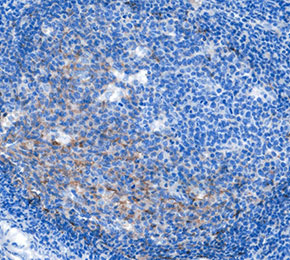 Immunohistochemical analysis of formalin fixed paraffin embedded human tonsil tissue with F9578 at 1:1000 dilution.