|
How to Cite 1. For In-Text Citation (Materials & Methods): 2. For Key Resources Table: |
||
|
Toll Free: (877) 796-6397 -- USA and Canada only -- |
Fax: +1-832-582-8590 Orders: +1-832-582-8158 |
Tech Support: +1-832-582-8158 Ext:3 Please provide your Order Number in the email. We strive to reply to |
Biological Description
| Specificity | CISD1/MitoNEET Antibody [M24F17] detects endogenous levels of total CISD1/MitoNEET protein. |
|---|---|
| Background | CDGSH iron–sulfur domain–containing protein 1 (CISD1), also known as mitoNEET, is an outer mitochondrial membrane protein of the NEET family that coordinates a redox‑active [2Fe–2S] cluster and regulates mitochondrial oxidative capacity, iron handling, and redox homeostasis. The protein contains a single C‑terminal transmembrane helix that anchors it in the outer mitochondrial membrane with the bulk of the protein facing the cytosol, and an N‑terminal cytosolic region organized around the characteristic CDGSH motif, which binds the [2Fe–2S] cluster in a labile, redox‑sensitive configuration stabilized by coordinating cysteine and histidine residues. This cluster confers redox activity and is susceptible to oxidative modification, allowing CISD1 to sense and respond to changes in the mitochondrial and cytosolic redox environment and to participate in transfer or sharing of iron–sulfur clusters with partner proteins. Functional data indicate that CISD1 modulates the maximal capacity for electron transport and oxidative phosphorylation by influencing the redox state of mitochondrial substrates and by regulating iron availability within mitochondria, which impacts respiratory chain function, reactive oxygen species production, and ATP generation. The protein is implicated in iron–sulfur cluster shuttling and iron trafficking, as its oxidized form can transfer the [2Fe–2S] cluster to apo‑acceptor proteins, while reduction destabilizes cluster binding, indicating a mechanism in which CISD1 operates as a redox‑regulated cluster donor and contributes to distribution of iron–sulfur cofactors under oxidative stress. Through these activities, CISD1 shapes mitochondrial iron and ROS homeostasis and thereby influences pathways such as ferroptosis, where iron‑dependent lipid peroxidation and mitochondrial dysfunction are key determinants of regulated necrotic cell death. In models of acute lung injury and other inflammatory settings, altered CISD1 expression correlates with changes in mitochondrial membrane potential, ATP levels, and ROS accumulation, and inhibition or reduction of CISD1 associates with protection against mitochondrial dysfunction and attenuation of ferroptotic and inflammatory markers, highlighting its regulatory position at the interface of metabolism, oxidative stress, and cell death pathways. Expression changes and functional modulation of CISD1 are reported in diverse disease contexts, including metabolic disorders, neurodegeneration, chronic obstructive pulmonary disease, and cancer, where its control over mitochondrial redox balance and iron handling influences cell survival, inflammatory activation, and proliferative behavior. |
Usage Information
| Application | WB, IHC, IF | Dilution |
|
||||||||||||
|---|---|---|---|---|---|---|---|---|---|---|---|---|---|---|---|
| Reactivity | Human, Mouse, Rat | ||||||||||||||
| Source | Rabbit Monoclonal Antibody | MW | 12 kDa | ||||||||||||
| Storage Buffer | PBS, pH 7.2+50% Glycerol+0.05% BSA+0.01% NaN3 | Storage (from the date of receipt) |
-20°C (avoid freeze-thaw cycles), 2 years | ||||||||||||
References
|
Application Data
WB
Validated by Selleck
-
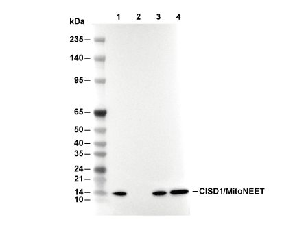 Lane 1: HeLa, Lane 2: HeLa (KO CISD1), Lane 3: 293T, Lane 4: Neuro-2a