|
How to Cite 1. For In-Text Citation (Materials & Methods): 2. For Key Resources Table: |
||
|
Toll Free: (877) 796-6397 -- USA and Canada only -- |
Fax: +1-832-582-8590 Orders: +1-832-582-8158 |
Tech Support: +1-832-582-8158 Ext:3 Please provide your Order Number in the email. We strive to reply to |
Biological Description
| Specificity | Cathepsin D (E5V4H) Rabbit mAb detects endogenous levels of total Cathepsin D protein. |
|---|---|
| Background | Cathepsin D belongs to the aspartyl protease family and functions primarily as a lysosomal enzyme responsible for intracellular protein degradation, with emerging roles in signaling and cell death regulation. It features a bilobal structure with two aspartic acid residues in the active site that coordinate substrate cleavage at acidic pH, maturing from an inactive proenzyme through autocatalytic processing into disulfide-linked heavy and light chains. Cathepsin D traffics from lysosomes to the cytosol during apoptosis, where both active and catalytically inactive forms enhance caspase activation and cytochrome c release independently of proteolysis, interacting directly with apoptotic machinery to amplify death receptor signaling; in cancer cells, secreted procathepsin D binds to cell surface receptors like M6PR, triggering proinvasive PAR1/EGFR pathways that promote matrix remodeling via hepsin degradation and uPAR activation, while cytosolic forms cleave Bcl-2 to relieve anti-apoptotic suppression. Cathepsin D, with its dual lysosomal-cytosolic action, serves as a versatile executor of programmed cell death and tissue remodeling. This makes it an ideal target for researchers investigating lysosomal membrane permeabilization or tumor microenvironment dynamics, where pH gradients critically regulate its enzymatic activity. It supports neuronal protein turnover and autophagy flux critical for synaptic maintenance and metabolic homeostasis, with ubiquitous expression enabling broad organelle quality control. Dysregulation through overexpression correlates with breast cancer metastasis and tamoxifen resistance, particularly in ER-positive tumors where high levels predict recurrence and distant spread. |
Usage Information
| Application | WB, IF, FCM | Dilution |
|
||||||
|---|---|---|---|---|---|---|---|---|---|
| Reactivity | Human, Mouse, Rat | ||||||||
| Source | Rabbit Monoclonal Antibody | MW | 46 kDa, 43 kDa, 28 kDa | ||||||
| Storage Buffer | PBS, pH 7.2+50% Glycerol+0.05% BSA+0.01% NaN3 | Storage (from the date of receipt) |
-20°C (avoid freeze-thaw cycles), 2 years | ||||||
References
|
Application Data
WB
Validated by Selleck
-
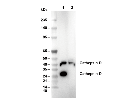 Lane 1: Mouse brain, Lane 2: Rat brain