|
How to Cite 1. For In-Text Citation (Materials & Methods): 2. For Key Resources Table: |
||
|
Toll Free: (877) 796-6397 -- USA and Canada only -- |
Fax: +1-832-582-8590 Orders: +1-832-582-8158 |
Tech Support: +1-832-582-8158 Ext:3 Please provide your Order Number in the email. We strive to reply to |
Biological Description
| Specificity | BNIP3 Antibody [A22K11] detects endogenous levels of total BNIP3 protein. |
|---|---|
| Background | BNIP3 (BCL2/adenovirus E1B 19 kDa–interacting protein 3) is an atypical BH3‑only member of the Bcl‑2 family that localizes predominantly to the outer mitochondrial membrane and functions as a hypoxia‑inducible regulator of mitochondrial integrity, cell death, and selective organelle autophagy. The protein contains an N‑terminal region with the BH3 motif that mediates interaction with anti‑apoptotic Bcl‑2 family members, a central intrinsically disordered segment, and a C‑terminal transmembrane domain that anchors BNIP3 in mitochondrial and, in some contexts, endoplasmic reticulum membranes and is required for homodimerization and pro‑death activity. BNIP3 expression increases strongly under hypoxic and metabolic stress conditions through direct transcriptional activation by HIF‑1α, and the protein then acts as a mitochondrial stress sensor that responds to redox changes by forming disulfide‑linked homodimers within the membrane, altering outer membrane permeability, dissipating mitochondrial membrane potential, and promoting release of death‑associated factors that drive caspase‑dependent or caspase‑independent cell death programs. BNIP3 also functions as a mitophagy receptor by using its LC3‑interacting region to recruit autophagosomal LC3 to damaged mitochondria and by cooperating with other quality‑control proteins such as SPATA18/MIEAP and BNIP3L/NIX, thereby coupling mitochondrial depolarization and accumulation of reactive oxygen species to selective engulfment and lysosomal degradation of dysfunctional mitochondria and contributing to maintenance of mitochondrial homeostasis under chronic stress. Dual localization of BNIP3 to mitochondria and endoplasmic reticulum allows initiation of distinct signaling outputs, with ER‑associated BNIP3 triggering ER‑selective autophagy (ER‑phagy) and modulating intracellular calcium partitioning between ER and mitochondria, which in turn influences susceptibility to apoptosis and necrosis and links BNIP3 activity to broader calcium‑dependent signaling networks. Post‑translational regulation, including phosphorylation events in the C‑terminal region by kinases such as JNK1/2, adjusts the balance between pro‑mitophagic and pro‑death functions by controlling dimer configuration and LC3 binding, thereby aligning BNIP3‑dependent mitochondrial clearance with hypoxic signaling and limiting excessive mitochondrial damage and cell loss in stress‑adapted tissues. Dysregulated BNIP3 expression and promoter methylation occur in multiple cancers, where loss or silencing of BNIP3 correlates with impaired hypoxia‑induced cell death and enhanced tumor survival in low‑oxygen microenvironments. |
Usage Information
| Application | WB, IP, IHC, IF | Dilution |
|
||||||||
|---|---|---|---|---|---|---|---|---|---|---|---|
| Reactivity | Human | ||||||||||
| Source | Rabbit Monoclonal Antibody | MW | 22 kDa | ||||||||
| Storage Buffer | PBS, pH 7.2+50% Glycerol+0.05% BSA+0.01% NaN3 | Storage (from the date of receipt) |
-20°C (avoid freeze-thaw cycles), 2 years | ||||||||
References
|
Application Data
WB
Validated by Selleck
-
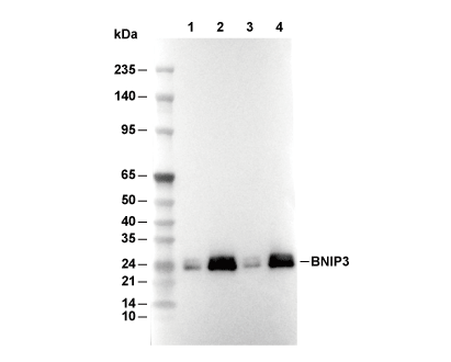 Lane 1: Hela, Lane 2: Hela (cobalt chloride, 100 μM, overnight), Lane 3: MCF7, Lane 4: MCF7 (cobalt chloride, 100 μM, overnight)