
|
How to Cite 1. For In-Text Citation (Materials & Methods): 2. For Key Resources Table: |
||
|
Toll Free: (877) 796-6397 -- USA and Canada only -- |
Fax: +1-832-582-8590 Orders: +1-832-582-8158 |
Tech Support: +1-832-582-8158 Ext:3 Please provide your Order Number in the email. We strive to reply to |
Biological Description
| Specificity | BACH2 Antibody (Rabbit mAb) [N18E12] detects endogenous levels of total BACH2 protein. |
|---|---|
| Background | BACH2, BTB and CNC homology 2, belongs to the BTB-basic leucine zipper family of transcriptional repressors, requiring heterodimerization with small Maf proteins such as MAFK to bind DNA at Maf recognition elements and repress target gene transcription, and it is predominantly expressed in B and T lymphocytes where it operates across two distinct branches of adaptive immunity. In germinal center B cells, BACH2 forms a complex with MAFK that binds chromatin at gene sets governing the germinal center response, and chromatin immunoprecipitation sequencing shows substantial overlap between BACH2 and BCL6 binding sites, including shared occupancy of regulatory sequences controlling PRDM1, the master driver of plasma cell differentiation. BCL6 and BACH2 cooperate through both physical and biochemical mechanisms, recruiting each other to shared DNA-binding sites or to proximal regulatory elements, and BCL6 additionally stabilizes BACH2 protein, so that the two factors' expression levels correlate positively within germinal center B cells; combined heterozygous loss of both factors, but not loss of either alone, produces a profound reduction in germinal center formation and accelerates premature plasmacytic differentiation, marked by elevated expression of Prdm1, Xbp1, and CD138, demonstrating that BCL6 and BACH2 function as a cooperative repressive unit rather than through independent, additive contributions. In the T cell compartment, BACH2 represses a broad network of effector-differentiation genes to stabilize regulatory T cell identity, restraining CD4-positive T cell differentiation toward inflammatory effector lineages while supporting Foxp3-dependent regulatory programming, and this repression extends to interleukin-2 receptor signaling: BACH2 directly represses CD25, the IL-2 receptor alpha chain, and this repression is required for the quiescence, survival, and long-term maintenance of resting regulatory T cells, with loss of BACH2 producing elevated CD25 and IL-2R signaling that only partially compensates for impaired Treg survival. BACH2-mediated CD25 repression additionally shapes T follicular regulatory cell development, coupling BACH2's transcriptional repressor activity to the balance between regulatory and effector states across multiple T cell subsets rather than to a single lineage decision. Across both B and T lymphocyte compartments, BACH2 functions as a rheostat restraining terminal effector or plasma cell differentiation in favor of quiescent, memory, or regulatory cell fates, and dysregulation of the BACH2-BCL6 balance or of BACH2-dependent Treg stabilization is implicated in autoimmune susceptibility and B-cell malignancy. |
Usage Information
| Application | WB, IP, FCM, ELISA | Dilution |
|
||||||
|---|---|---|---|---|---|---|---|---|---|
| Reactivity | Human, Mouse | ||||||||
| Source | Rabbit Monoclonal Antibody | MW | 93 kDa | ||||||
| Storage Buffer | PBS, pH 7.2+50% Glycerol+0.05% BSA+0.01% NaN3 | Storage (from the date of receipt) |
-20°C (avoid freeze-thaw cycles), 2 years | ||||||
| WB |
Experimental Protocol:
Sample preparation
1. Tissue: Lyse the tissue sample by adding an appropriate volume of ice-cold RIPA/NP-40 Lysis Buffer (containing Protease Inhibitor Cocktail),and homogenize the tissue at a low temperature or lyse it by sonication on ice, then incubate on ice for 30 minutes. 2. Adherent cell: Aspirate the culture medium and wash the cells with ice-cold PBS twice. Lyse the cells by adding an appropriate volume of RIPA/NP-40 Lysis Buffer (containing Protease Inhibitor Cocktail) , sonicate to lyse the cells, and incubate on ice for 30 minutes. 3. Suspension cell: Transfer the culture medium to a pre-cooled centrifuge tube. Centrifuge and aspirate the supernatant. Wash the cells with ice-cold PBS twice. Lyse the cells by adding an appropriate volume of RIPA/NP-40 Lysis Buffer (containing Protease Inhibitor Cocktail) , sonicate to lyse the cells, and incubate on ice for 30 minutes. 4. Place the lysate into a pre-cooled microcentrifuge tube. Centrifuge at 4°C for 15 min. Collect the supernatant;
5. Remove a small volume of lysate to determine the protein concentration;
6. Combine the lysate with protein loading buffer. Boil 20 µL sample under 95-100°C for 5 min. Centrifuge for 5 min after cool down on ice.
Electrophoretic separation
1. According to the concentration of extracted protein, load appropriate amount of protein sample and marker onto SDS-PAGE gels for electrophoresis. Recommended separating gel (lower gel) concentration: 5%. Reference Table for Selecting SDS-PAGE Separation Gel Concentrations 2. Power up 80V for 30 minutes. Then the power supply is adjusted (110 V~150 V), the Marker is observed, and the electrophoresis can be stopped when the indicator band of the predyed protein Marker where the protein is located is properly separated. (Note that the current should not be too large when electrophoresis, too large current (more than 150 mA) will cause the temperature to rise, affecting the result of running glue. If high currents cannot be avoided, an ice bath can be used to cool the bath.)
Transfer membrane
1. Take out the converter, soak the clip and consumables in the pre-cooled converter;
2. Activate PVDF membrane with methanol for 1 min and rinse with transfer buffer;
3. Install it in the order of "black edge of clip - sponge - filter paper - filter paper - glue -PVDF membrane - filter paper - filter paper - sponge - white edge of clip"; 4. The protein was electrotransferred to PVDF membrane. ( 0.45 µm PVDF membrane is recommended ) Reference Table for Selecting PVDF Membrane Pore Size Specifications Recommended conditions for wet transfer: 200 mA, 120 min. ( Note that the transfer conditions can be adjusted according to the protein size. For high-molecular-weight proteins, a higher current and longer transfer time are recommended. However, ensure that the transfer tank remains at a low temperature to prevent gel melting.)
Block
1. After electrotransfer, wash the film with TBST at room temperature for 5 minutes;
2. Incubate the film in the blocking solution for 1 hour at room temperature;
3. Wash the film with TBST for 3 times, 5 minutes each time.
Antibody incubation
1. Use primary antibody dilution buffer to prepare the primary antibody working liquid (recommended dilution ratio for primary antibody 1:1000), gently shake and incubate with the film at 4°C overnight; 2. Wash the film with TBST 3 times, 5 minutes each time;
3. Add the secondary antibody to the blocking solution and incubate with the film gently at room temperature for 1 hour;
4. After incubation, wash the film with TBST 3 times for 5 minutes each time.
Antibody staining
1. Add the prepared ECL luminescent substrate (or select other color developing substrate according to the second antibody) and mix evenly;
2. Incubate with the film for 1 minute, remove excess substrate (keep the film moist), wrap with plastic film, and expose in the imaging system.
|
References
|
Application Data
WB
Validated by Selleck
-
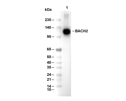 Lane 1: Ramos