|
Toll Free: (877) 796-6397 -- USA and Canada only -- |
Fax: +1-832-582-8590 Orders: +1-832-582-8158 |
Tech Support: +1-832-582-8158 Ext:3 Please provide your Order Number in the email. |
Biological Description
| Specificity | Anti-Poly/Mono-ADP Ribose Rabbit Antibody [A22J18] recognizes endogenous levels of ADP ribosylated proteins and does not cross-react with other post translational modifications. |
|---|---|
| Background | Poly(ADP-ribose) (PAR) and mono(ADP-ribose) (MAR) are reversible post-translational modifications mediated by ADP-ribosyltransferases, particularly members of the poly(ADP-ribose) polymerase (PARP) family. MAR refers to the covalent addition of a single ADP-ribose unit—transferred from NAD⁺—to specific amino acid residues (commonly aspartate, glutamate, or lysine) on target proteins. In contrast, PAR consists of two or more ADP-ribose units linked through ribose–ribose glycosidic bonds, forming linear or branched chains. Structurally, each ADP-ribose unit contributes ~0.5 kDa, making PAR a large, highly negatively charged and flexible polymer capable of scaffolding diverse protein assemblies. PARP1, the most abundant and catalytically active nuclear PARP, is rapidly activated by DNA strand breaks and stress signals, catalyzing PARylation to recruit and organize DNA repair complexes. MARylation and PARylation play distinct cellular roles—MAR often regulates individual protein activity or interactions, while PAR serves broader functions as a dynamic structural element in processes such as DNA repair, chromatin remodeling, stress granule formation, mitotic spindle assembly, and nucleolar integrity. The balance between synthesis by PARPs and degradation by enzymes like poly(ADP-ribose) glycohydrolase (PARG) and macrodomain hydrolases is critical for maintaining cellular homeostasis. |
Usage Information
| Application | WB, IF | Dilution |
|
||||
|---|---|---|---|---|---|---|---|
| Reactivity | All | ||||||
| Source | Rabbit | MW | |||||
| Storage Buffer | PBS, pH 7.2+50% Glycerol+0.05% BSA+0.01% NaN₃ | Storage (from the date of receipt) |
-20°C (avoid freeze-thaw cycles), 2 years | ||||
| IF |
Experimental Protocol:
Specimen Preparation
1. Aspirate liquid, then cover cells to a depth of 2–3 mm with 4% Paraformaldehyde diluted in 1X PBS.
NOTE: Paraformaldehyde is toxic, use only in a fume hood.
2. Fix cells for 15 min at room temperature.
3. Aspirate fixative, rinse three times in 1X PBS for 5 min each.
4. Proceed with Immunostaining.
Immunostaining
1. Add theblocking buffer and incubate for 60 min at RT.
2. Prepare primary antibody diluent in antibody dilution buffer as recommended .
3. Aspirate blocking solution, apply diluted primary antibody.
4. Incubate overnight at 4°C.
5. Rinse three times in 1X PBS for 5 min each.
6. Incubate specimens in fluorochrome-conjugated secondary antibody diluted in antibody dilution buffer for 1–2 hr at room temperature in the dark.
7. Rinse three times in 1X PBS for 5 min each.
8. Mount slides usingmounting medium with DAPI and cover with coverslips.
9. For best results, allow mountant to cure overnight at room temperature. For long-term storage, store slides flat at 59°C protected from light.
|
References
|
Application Data
IF
Validated by Selleck
-
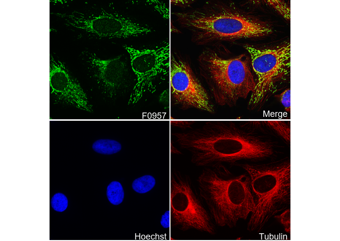 Immunofluorescent analysis of Hela cells using F0957 (green, 1:12000 ), Hoechst (blue) and tubulin (Red).