|
Toll Free: (877) 796-6397 -- USA and Canada only -- |
Fax: +1-832-582-8590 Orders: +1-832-582-8158 |
Tech Support: +1-832-582-8158 Ext:3 Please provide your Order Number in the email. |
Biological Description
| Specificity | Anti-Perforin Mouse Antibody [P20D17] recognizes endogenous levels of total Perforin protein. |
|---|---|
| Background | Perforin is a pore-forming glycoprotein mainly produced by cytotoxic lymphocytes such as natural killer (NK) cells and cytotoxic T lymphocytes (CTLs). It belongs to the MACPF (membrane attack complex/perforin) family and consists of domains critical for its function: the MACPF domain responsible for pore formation, a calcium-dependent C2 domain mediating membrane binding, and an EGF-like domain providing structural flexibility. Once secreted into the immunological synapse formed between the cytotoxic cell and target (virus-infected or malignant) cell, perforin oligomerizes and inserts into the target cell membrane, forming transmembrane pores approximately 20 nm in diameter. These pores allow entry of pro-apoptotic granzymes into the cytosol, which trigger apoptosis of the target cell. Key residues such as R298 are important for oligomerization and pore formation. Perforin-mediated pore formation is essential for immune surveillance and elimination of abnormal cells, and deficiencies or mutations in perforin cause severe immunodeficiency disorders like familial hemophagocytic lymphohistiocytosis (FHL). Perforin also plays roles in immune regulation and is implicated when cytotoxic function is disrupted or dysregulated in various diseases. |
Usage Information
| Application | IHC | Dilution |
|
||
|---|---|---|---|---|---|
| Reactivity | Human | ||||
| Source | Mouse | MW | 70 kDa | ||
| Storage Buffer | PBS, pH 7.2+50% Glycerol+0.05% BSA+0.01% NaN3 | Storage (from the date of receipt) |
-20°C (avoid freeze-thaw cycles), 2 years | ||
| IHC |
Experimental Protocol:
Deparaffinization/Rehydration
1. Deparaffinize/hydrate sections:
2. Incubate sections in three washes of xylene for 5 min each.
3. Incubate sections in two washes of 100% ethanol for 10 min each.
4. Incubate sections in two washes of 95% ethanol for 10 min each.
5. Wash sections two times in dH2O for 5 min each.
6.Antigen retrieval: For Citrate: Heat slides in a microwave submersed in 1X citrate unmasking solution until boiling is initiated; continue with 10 min at a sub-boiling temperature (95°-98°C). Cool slides on bench top for 30 min.
Staining
1. Wash sections in dH2O three times for 5 min each.
2. Incubate sections in 3% hydrogen peroxide for 10 min.
3. Wash sections in dH2O two times for 5 min each.
4. Wash sections in wash buffer for 5 min.
5. Block each section with 100–400 µl of blocking solution for 1 hr at room temperature.
6. Remove blocking solution and add 100–400 µl primary antibody diluent in to each section. Incubate overnight at 4°C.
7. Remove antibody solution and wash sections with wash buffer three times for 5 min each.
8. Cover section with 1–3 drops HRPas needed. Incubate in a humidified chamber for 30 min at room temperature.
9. Wash sections three times with wash buffer for 5 min each.
10. Add DAB Chromogen Concentrate to DAB Diluent and mix well before use.
11. Apply 100–400 µl DAB to each section and monitor closely. 1–10 min generally provides an acceptable staining intensity.
12. Immerse slides in dH2O.
13. If desired, counterstain sections with hematoxylin.
14. Wash sections in dH2O two times for 5 min each.
15. Dehydrate sections: Incubate sections in 95% ethanol two times for 10 sec each; Repeat in 100% ethanol, incubating sections two times for 10 sec each; Repeat in xylene, incubating sections two times for 10 sec each.
16. Mount sections with coverslips and mounting medium.
|
References
|
Application Data
IHC
Validated by Selleck
-
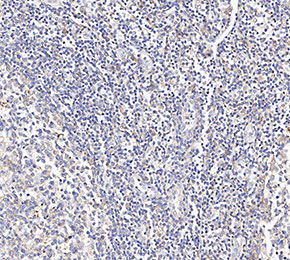 Immunohistochemical analysis of formalin fixed paraffin embedded human tonsils tissue with F2444 at 1:100 dilution.
IHC
Validated by Selleck
-
Immunohistochemical analysis of formalin fixed paraffin embedded human tonsils tissue with F2444 at 1:100 dilution.